

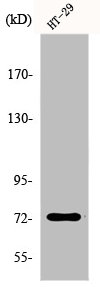
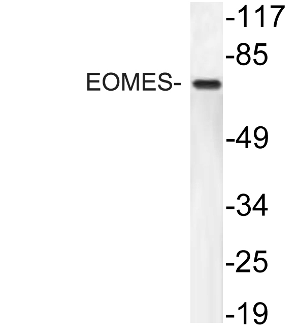
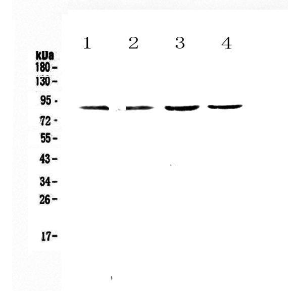
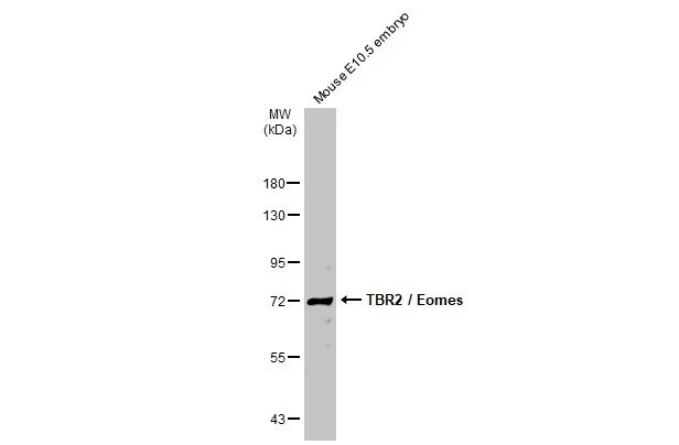
Western Blot analysis of HepG2 cells using EOMES Polyclonal Antibody
Western Blot analysis of HepG2 cells using EOMES Polyclonal Antibody
EOMES Antibody
CSB-PA002351
ApplicationsWestern Blot, ELISA
Product group Antibodies
ReactivityHuman, Mouse
TargetEOMES
Overview
- SupplierCusabio
- Product NameEOMES Antibody
- Delivery Days Customer20
- ApplicationsWestern Blot, ELISA
- CertificationResearch Use Only
- ClonalityPolyclonal
- ConjugateUnconjugated
- Gene ID8320
- Target nameEOMES
- Target descriptioneomesodermin
- Target synonymsTBR2, eomesodermin homolog, T-box brain protein 2
- HostRabbit
- IsotypeIgG
- Protein IDO95936
- Protein NameEomesodermin homolog
- ReactivityHuman, Mouse
- Storage Instruction-20°C or -80°C
- UNSPSC41116161
Related products
Product group Antibodies
Anti-EOMES AntibodyA99114
ApplicationsWestern Blot, ELISA
ReactivityHuman, Mouse
- SizePrice
Product group Antibodies
Anti-TBR2/Eomes Antibody Picoband(r)A00992-1-CARRIER-FREE
ApplicationsWestern Blot, ELISA
ReactivityHuman, Mouse, Rat
TargetEOMES
- SizePrice
Product group Antibodies

Eomesodermin / EOMES Antibody (HRP)LS-C674006
ReactivityHuman
TargetEOMES
- SizePrice
Product group Antibodies
EOMES Polyclonal AntibodyCAC15095
ApplicationsWestern Blot, ELISA, ImmunoHistoChemistry
ReactivityRat
TargetEOMES
- SizePrice
Product group Antibodies
TBR2 / Eomes antibodyGTX132776
ApplicationsWestern Blot, ImmunoHistoChemistry, ImmunoHistoChemistry Paraffin
ReactivityHuman, Mouse
TargetEOMES
- SizePrice

Product group Antibodies
Anti-EOMES AntibodyHPA028896
ApplicationsImmunoHistoChemistry
ReactivityHuman
TargetEOMES
- SizePrice