

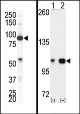
(LEFT)Western blot analysis of anti-EphA4 Pab in NCI-H460 cell lysate. EphA4 (arrow) was detected using purified Pab. Secondary HRP-anti-rabbit was used for signal visualization with chemiluminescence. (RIGHT)Western blot analysis of EPHA4 (arrow) using EphA4 Antibody (C-term). 293 cell lysates (2 ug/lane) either nontransfected (Lane 1) or transiently transfected with the EPHA4 gene (Lane 2)
(LEFT)Western blot analysis of anti-EphA4 Pab in NCI-H460 cell lysate. EphA4 (arrow) was detected using purified Pab. Secondary HRP-anti-rabbit was used for signal visualization with chemiluminescence. (RIGHT)Western blot analysis of EPHA4 (arrow) using EphA4 Antibody (C-term). 293 cell lysates (2 ug/lane) either nontransfected (Lane 1) or transiently transfected with the EPHA4 gene (Lane 2)
Eph receptor A4 (EPHA4) (C-term) Rabbit Polyclonal Antibody
AP14279PU-N
ApplicationsWestern Blot, ImmunoHistoChemistry
Product group Antibodies
ReactivityHuman
TargetEPHA4
Overview
- SupplierOriGene
- Product NameEph receptor A4 (EPHA4) (C-term) Rabbit Polyclonal Antibody
- Delivery Days Customer14
- ApplicationsWestern Blot, ImmunoHistoChemistry
- CertificationResearch Use Only
- ClonalityPolyclonal
- Gene ID2043
- Target nameEPHA4
- Target descriptionEPH receptor A4
- Target synonymsEK8, HEK8, SEK, TYRO1, ephrin type-A receptor 4, EPH-like kinase 8, TYRO1 protein tyrosine kinase, receptor protein-tyrosine kinase HEK8, tyrosine-protein kinase TYRO1, tyrosine-protein kinase receptor SEK
- HostRabbit
- Protein IDP54764
- Protein NameEphrin type-A receptor 4
- Scientific DescriptionEph receptor A4 (EPHA4) (C-term) rabbit polyclonal antibody, Purified
- ReactivityHuman
- Storage Instruction-20°C,2°C to 8°C
- UNSPSC12352203
MSDS
Related products
Product group Antibodies

EPHA4 (Phospho-Tyr596) AntibodyABX012736
ApplicationsWestern Blot, ELISA
- SizePrice
Product group Antibodies
Anti-EPHA4 Antibody144-08346
ApplicationsWestern Blot
ReactivityHuman, Mouse, Rat
TargetEPHA4
- SizePrice
![Non-transfected (–) and transfected (+) 293T whole cell extracts (30 μg) were separated by 7.5% SDS-PAGE, and the membrane was blotted with EphA4 antibody [N3C2], Internal (GTX104109) diluted at 1:5000. The HRP-conjugated anti-rabbit IgG antibody (GTX213110-01) was used to detect the primary antibody.](https://www.genetex.com/upload/website/prouct_img/normal/GTX104109/GTX104109_40583_20180727_WB_B_w_23060120_399.webp)
Product group Antibodies
EphA4 antibody [N3C2], InternalGTX104109
ApplicationsWestern Blot
ReactivityHuman, Mouse, Rat
TargetEPHA4
- SizePrice
Product group Antibodies
EPHA4 Polyclonal AntibodyCAC12899
ApplicationsImmunoFluorescence, ELISA, ImmunoHistoChemistry
TargetEPHA4
- SizePrice
Product group Antibodies
ApplicationsImmunoFluorescence, ELISA, ImmunoCytoChemistry, ImmunoHistoChemistry, ImmunoHistoChemistry Frozen, ImmunoHistoChemistry Paraffin
ReactivityCanine, Chicken, Equine, Human, Mouse, Porcine, Rabbit, Rat
TargetEPHA4
- SizePrice
Product group Antibodies
Anti-EPHA4 AntibodyA46071
ApplicationsImmunoHistoChemistry
ReactivityHuman, Mouse
- SizePrice
Product group Antibodies
EPHA4 AntibodyCSB-PA007724LA01HU
ApplicationsImmunoFluorescence, ELISA, ImmunoHistoChemistry
ReactivityHuman
TargetEPHA4
- SizePrice
Product group Antibodies

ApplicationsWestern Blot
ReactivityChicken, Human, Mouse
TargetEPHA4
- SizePrice