

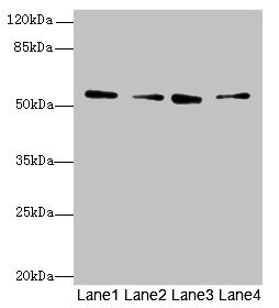
Western blot All lanes: ERO1B antibody at 3.2microg/ml Lane 1: MCF-7 whole cell lysate Lane 2: U251 whole cell lysate Lane 3: Mouse liver tissue Lane 4: U87 whole cell lysate Secondary Goat polyclonal to rabbit IgG at 1/10000 dilution Predicted band size: 54, 17 kDa Observed band size: 54 kDa


Western blot All lanes: ERO1B antibody at 3.2microg/ml Lane 1: MCF-7 whole cell lysate Lane 2: U251 whole cell lysate Lane 3: Mouse liver tissue Lane 4: U87 whole cell lysate Secondary Goat polyclonal to rabbit IgG at 1/10000 dilution Predicted band size: 54, 17 kDa Observed band size: 54 kDa
ERO1B Antibody
CSB-PA774834LA01HU
ApplicationsWestern Blot, ELISA, ImmunoHistoChemistry
Product group Antibodies
ReactivityHuman, Mouse
TargetERO1B
Overview
- SupplierCusabio
- Product NameERO1B Antibody
- Delivery Days Customer20
- ApplicationsWestern Blot, ELISA, ImmunoHistoChemistry
- CertificationResearch Use Only
- ClonalityPolyclonal
- ConjugateUnconjugated
- Gene ID56605
- Target nameERO1B
- Target descriptionendoplasmic reticulum oxidoreductase 1 beta
- Target synonymsERO1LB, Ero1beta, ERO1-like protein beta, ERO1-L-beta, ERO1-like beta, endoplasmic oxidoreductin-1-like protein B, endoplasmic reticulum oxidoreductase beta, endoplasmic reticulum oxidoreductin-1-like protein B, oxidoreductin-1-L-beta
- HostRabbit
- IsotypeIgG
- Protein IDQ86YB8
- Protein NameERO1-like protein beta
- Scientific DescriptionOxidoreductase involved in disulfide bond formation in the endoplasmic reticulum. Efficiently reoxidizes P4HB/PDI, the enzyme catalyzing protein disulfide formation, in order to allow P4HB to sustain additional rounds of disulfide formation. Other protein disulfide isomerase family members can also be reoxidized, but at lower rates compared to P4HB, including PDIA2 (50% of P4HB reoxidation rate), as well as PDIA3, PDIA4, PDIA6 and NXNDC12 (<10%). Following P4HB reoxidation, passes its electrons to molecular oxygen via FAD, leading to the production of reactive oxygen species (ROS) in the cell. May be involved in oxidative proinsulin folding in pancreatic cells, hence may play a role in glucose homeostasis.
- ReactivityHuman, Mouse
- Storage Instruction-20°C or -80°C
- UNSPSC41116161
Related products
Product group Antibodies
References
ERO1LB Polyclonal AntibodyBS-14627R
ApplicationsImmunoFluorescence, Western Blot, ELISA, ImmunoCytoChemistry, ImmunoHistoChemistry, ImmunoHistoChemistry Frozen, ImmunoHistoChemistry Paraffin
ReactivityGuinea Pig, Human, Mouse, Rabbit, Rat
TargetERO1B
- SizePrice
Product group Antibodies
Ero1B Polyclonal AntibodyCAC09445
ApplicationsWestern Blot, ELISA, ImmunoHistoChemistry
ReactivityMouse
TargetERO1B
- SizePrice
Product group Antibodies

ERO1LB AntibodyLS-C332714
ApplicationsWestern Blot
ReactivityHuman, Mouse
TargetERO1B
- SizePrice
Product group Antibodies
Anti-ERO1B AntibodyHPA028085
ApplicationsWestern Blot, ImmunoHistoChemistry
ReactivityHuman
TargetERO1B
- SizePrice
Product group Antibodies
ERO1LB antibodyGTX53598
ApplicationsFlow Cytometry, Western Blot, ImmunoHistoChemistry, ImmunoHistoChemistry Paraffin
ReactivityHuman, Mouse
TargetERO1B
- SizePrice