

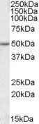
WB analysis of Jurkat lysate using GTX47560 EVL antibody, Internal. Dilution : 1μg/ml Loading : 35μg protein in RIPA buffer
WB analysis of Jurkat lysate using GTX47560 EVL antibody, Internal. Dilution : 1μg/ml Loading : 35μg protein in RIPA buffer
EVL antibody, Internal
GTX47560
ApplicationsWestern Blot
Product group Antibodies
ReactivityHuman
TargetEVL
Overview
- SupplierGeneTex
- Product NameEVL antibody, Internal
- Delivery Days Customer7
- Application Supplier NoteWB: 1-3microg/ml. *Optimal dilutions/concentrations should be determined by the researcher.Not tested in other applications.
- ApplicationsWestern Blot
- CertificationResearch Use Only
- ClonalityPolyclonal
- Concentration0.50 mg/ml
- ConjugateUnconjugated
- Gene ID51466
- Target nameEVL
- Target descriptionEnah/Vasp-like
- Target synonymsRNB6, ena/VASP-like protein, ena/vasodilator-stimulated phosphoprotein-like, epididymis secretory sperm binding protein
- HostGoat
- IsotypeIgG
- Protein IDQ9UI08
- Protein NameEna/VASP-like protein
- ReactivityHuman
- Storage Instruction-20°C or -80°C,2°C to 8°C
- UNSPSC41116161
Datasheet
Related products
Product group Antibodies
EVL AntibodyCSB-PA007869GA01HU
ApplicationsWestern Blot, ELISA
ReactivityHuman, Mouse, Rat
TargetEVL
- SizePrice

Product group Antibodies
Anti-EVL Antibody Picoband(r)A02568-2-CARRIER-FREE
ApplicationsFlow Cytometry, Western Blot, ELISA, ImmunoHistoChemistry
ReactivityHuman
TargetEVL
- SizePrice
Product group Antibodies
Anti-EVL AntibodyHPA018849
ApplicationsWestern Blot, ImmunoHistoChemistry
ReactivityHuman
TargetEVL
- SizePrice
Product group Antibodies

EVL AntibodyLS-C661530
ApplicationsFlow Cytometry, ImmunoFluorescence, ImmunoPrecipitation, Western Blot, ELISA, ImmunoHistoChemistry
ReactivityHuman
TargetEVL
- SizePrice