

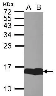
Sample (30 μg of whole cell lysate) A: whole zebrafish B: Zebrafish brain 15% SDS PAGE GTX121467 diluted at 1:1000
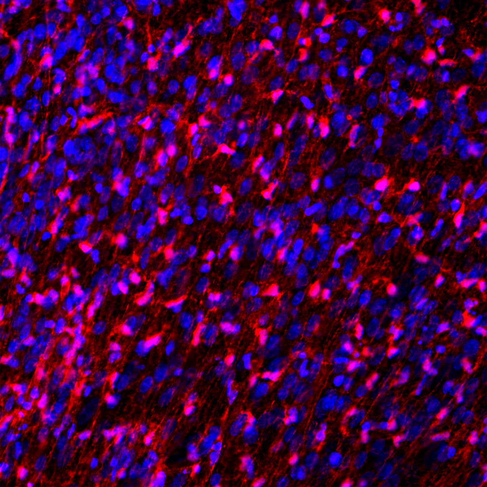
![FABP7 antibody [N1C3] detects FABP7 protein expression by immunohistochemical analysis.
Sample: Frozen sectioned E13.5 Rat brain.
Green: FABP7 protein stained by FABP7 antibody [N1C3] (GTX121467) diluted at 1:250.
Red: beta Tubulin 3/ TUJ1, a mature neuron marker, stained by beta Tubulin 3/ TUJ1 antibody [GT11710] (GTX631836) diluted at 1:500.
Blue: Fluoroshield with DAPI (GTX30920).](https://www.genetex.com/upload/website/prouct_img/normal/GTX121467/GTX121467_40492_20160921_IHC-Fr_w_23060519_924.webp "FABP7 antibody [N1C3] detects FABP7 protein expression by immunohistochemical analysis.
Sample: Frozen sectioned E13.5 Rat brain.
Green: FABP7 protein stained by FABP7 antibody [N1C3] (GTX121467) diluted at 1:250.
Red: beta Tubulin 3/ TUJ1, a mature neuron marker, stained by beta Tubulin 3/ TUJ1 antibody [GT11710] (GTX631836) diluted at 1:500.
Blue: Fluoroshield with DAPI (GTX30920).")
 A: NT2D1 B: IMR32 15% SDS PAGE GTX121467 diluted at 1:1000")
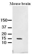
![Various tissue extracts (50 μg) were separated by 15% SDS-PAGE, and the membrane was blotted with FABP7 antibody [N1C3] (GTX121467) diluted at 1:1000.](https://www.genetex.com/upload/website/prouct_img/normal/GTX121467/GTX121467_40492_20160825_WB_M_R_w_23060519_571.webp "Various tissue extracts (50 μg) were separated by 15% SDS-PAGE, and the membrane was blotted with FABP7 antibody [N1C3] (GTX121467) diluted at 1:1000.")
![FABP7 antibody [N1C3] detects FABP7 protein by immunofluorescent analysis. Sample: DIV9 rat E18 primary hippocampal neuron and glia cells were fixed in 4% paraformaldehyde at RT for 15 min. Green: FABP7 stained by FABP7 antibody [N1C3] (GTX121467) diluted at 1:500. Red: beta Tubulin 3/ Tuj1, stained by beta Tubulin 3/ Tuj1 antibody [GT11710] (GTX631836) diluted at 1:500. Blue: Fluoroshield with DAPI (GTX30920).](https://www.genetex.com/upload/website/prouct_img/normal/GTX121467/GTX121467_40492_20181004_ICC_IF_R_w_23060519_937.webp "FABP7 antibody [N1C3] detects FABP7 protein by immunofluorescent analysis. Sample: DIV9 rat E18 primary hippocampal neuron and glia cells were fixed in 4% paraformaldehyde at RT for 15 min. Green: FABP7 stained by FABP7 antibody [N1C3] (GTX121467) diluted at 1:500. Red: beta Tubulin 3/ Tuj1, stained by beta Tubulin 3/ Tuj1 antibody [GT11710] (GTX631836) diluted at 1:500. Blue: Fluoroshield with DAPI (GTX30920).")
Sample (30 μg of whole cell lysate) A: whole zebrafish B: Zebrafish brain 15% SDS PAGE GTX121467 diluted at 1:1000
FABP7 antibody [N1C3]
GTX121467
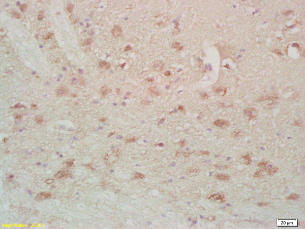
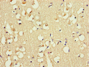
ApplicationsImmunoFluorescence, Western Blot, ImmunoCytoChemistry, ImmunoHistoChemistry, ImmunoHistoChemistry Frozen
Product group Antibodies
ReactivityHuman, Mouse, Rat, Zebra Fish
TargetFABP7
Overview
- SupplierGeneTex
- Product NameFABP7 antibody [N1C3]
- Delivery Days Customer9
- Application Supplier NoteWB: 1:500-1:3000. ICC/IF: 1:100-1:1000. IHC-Fr: 1:100-1:1000. *Optimal dilutions/concentrations should be determined by the researcher.Not tested in other applications.
- ApplicationsImmunoFluorescence, Western Blot, ImmunoCytoChemistry, ImmunoHistoChemistry, ImmunoHistoChemistry Frozen
- CertificationResearch Use Only
- ClonalityPolyclonal
- Concentration1 mg/ml
- ConjugateUnconjugated
- Gene ID2173
- Target nameFABP7
- Target descriptionfatty acid binding protein 7
- Target synonymsB-FABP, BLBP, FABPB, MRG, fatty acid-binding protein, brain, brain lipid-binding protein, brain-type fatty acid-binding protein, hypothetical protein DKFZp547J2313, mammary-derived growth inhibitor-related
- HostRabbit
- IsotypeIgG
- Protein IDO15540
- Protein NameFatty acid-binding protein, brain
- Scientific DescriptionThe protein encoded by this gene is a brain fatty acid binding protein. Fatty acid binding proteins (FABPs) are a family of small, highly conserved, cytoplasmic proteins that bind long-chain fatty acids and other hydrophobic ligands. FABPs are thought to play roles in fatty acid uptake, transport, and metabolism. [provided by RefSeq]
- ReactivityHuman, Mouse, Rat, Zebra Fish
- Storage Instruction-20°C or -80°C,2°C to 8°C
- UNSPSC41116161
Datasheet
Related products
Product group Antibodies
Anti-FABP7 AntibodyA104339
ApplicationsImmunoFluorescence, Western Blot, ImmunoCytoChemistry
ReactivityHuman, Mouse, Rat
- SizePrice
Product group Antibodies
Anti-FABP7 Antibody144-11604
ApplicationsWestern Blot
ReactivityHuman, Mouse, Rat
TargetFABP7
- SizePrice

Product group Antibodies
Anti-FABP7 AntibodyAMAB90595
ApplicationsWestern Blot, ImmunoHistoChemistry
ReactivityHuman
TargetFABP7
- SizePrice
Product group Antibodies
FABP7 Polyclonal AntibodyBS-2135R
ApplicationsImmunoFluorescence, ELISA, ImmunoCytoChemistry, ImmunoHistoChemistry, ImmunoHistoChemistry Frozen, ImmunoHistoChemistry Paraffin
ReactivityHuman, Mouse, Rat
TargetFABP7
- SizePrice
Product group Antibodies
FABP7 AntibodyCSB-PA007956LA01HU
ApplicationsImmunoFluorescence, ELISA, ImmunoHistoChemistry
ReactivityHuman
TargetFABP7
- SizePrice
Product group Antibodies

Goat anti-B-FABP/ BLBPEB09274
ApplicationsELISA, ImmunoHistoChemistry
ReactivityBovine, Human, Porcine
TargetFABP7
- SizePrice
Product group Antibodies
ApplicationsWestern Blot, ELISA, ImmunoCytoChemistry, ImmunoHistoChemistry, ImmunoHistoChemistry Frozen, ImmunoHistoChemistry Paraffin
ReactivityBovine, Mouse, Porcine, Rat
TargetFABP7
- SizePrice
Product group Antibodies

FABP7 / BLBP / MRG AntibodyLS-C403991
ApplicationsWestern Blot, ELISA, ImmunoHistoChemistry
ReactivityHuman, Mouse, Rat
TargetFABP7
- SizePrice
Product group Antibodies
FABP7 antibody [AT1D1]GTX50031
ApplicationsWestern Blot, ELISA
ReactivityHuman, Mouse
TargetFABP7
- SizePrice