

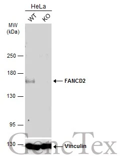
Wild-type (WT) and FANCD2 knockout (KO) HeLa cell extracts (30 μg) were separated by 5% SDS-PAGE, and the membrane was blotted with FANCD2 antibody [103] (GTX70299) diluted at 1:500. The HRP-conjugated anti-mouse IgG antibody (GTX213111-01) was used to detect the primary antibody.
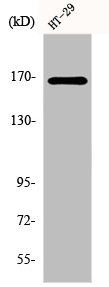
![Various whole cell extracts (30 μg) were separated by 5% SDS-PAGE, and the membrane was blotted with FANCD2 antibody [103] (GTX70299) diluted at 1:1000. The HRP-conjugated anti-mouse IgG antibody (GTX213111-01) was used to detect the primary antibody, and the signal was developed with Trident ECL plus-Enhanced.](https://www.genetex.com/upload/website/prouct_img/normal/GTX70299/GTX70299_41330_20211029_WB_w_23061221_509.webp "Various whole cell extracts (30 μg) were separated by 5% SDS-PAGE, and the membrane was blotted with FANCD2 antibody [103] (GTX70299) diluted at 1:1000. The HRP-conjugated anti-mouse IgG antibody (GTX213111-01) was used to detect the primary antibody, and the signal was developed with Trident ECL plus-Enhanced.")
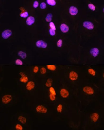
![FANCD2 antibody [103] detects FANCD2 protein at nucleus by immunofluorescent analysis. Sample: HeLa cells were fixed in 4% paraformaldehyde at RT for 15 min. Green: FANCD2 stained by FANCD2 antibody [103] (GTX70299) diluted at 1:500. Red: alpha Tubulin, a cytoskeleton marker, stained by alpha Tubulin antibody (GTX102079) diluted at 1:1000.](https://www.genetex.com/upload/website/prouct_img/normal/GTX70299/GTX70299_44489_20220121_ICC_IF_w_23061221_845.webp "FANCD2 antibody [103] detects FANCD2 protein at nucleus by immunofluorescent analysis. Sample: HeLa cells were fixed in 4% paraformaldehyde at RT for 15 min. Green: FANCD2 stained by FANCD2 antibody [103] (GTX70299) diluted at 1:500. Red: alpha Tubulin, a cytoskeleton marker, stained by alpha Tubulin antibody (GTX102079) diluted at 1:1000.")
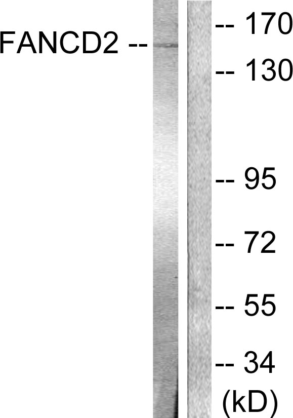
![FANCD2 antibody [103]](https://www.genetex.com/upload/website/prouct_img/normal/GTX70299/FANCD2-antibody-103-GTX70299-WB-1_w_23061221_113.webp "FANCD2 antibody [103]")
Wild-type (WT) and FANCD2 knockout (KO) HeLa cell extracts (30 μg) were separated by 5% SDS-PAGE, and the membrane was blotted with FANCD2 antibody [103] (GTX70299) diluted at 1:500. The HRP-conjugated anti-mouse IgG antibody (GTX213111-01) was used to detect the primary antibody.
FANCD2 antibody [103]
GTX70299
ApplicationsImmunoFluorescence, Western Blot, ELISA, ImmunoCytoChemistry
Product group Antibodies
ReactivityHuman
TargetFANCD2
Overview
- SupplierGeneTex
- Product NameFANCD2 antibody [103]
- Delivery Days Customer9
- Application Supplier NoteWB: 1:500-1:3000. *Optimal dilutions/concentrations should be determined by the researcher.Not tested in other applications.
- ApplicationsImmunoFluorescence, Western Blot, ELISA, ImmunoCytoChemistry
- CertificationResearch Use Only
- ClonalityMonoclonal
- Clone ID103
- Concentration1 mg/ml
- ConjugateUnconjugated
- Gene ID2177
- Target nameFANCD2
- Target descriptionFA complementation group D2
- Target synonymsFA-D2, FA4, FACD, FAD, FAD2, FANCD, Fanconi anemia group D2 protein, Fanconi anemia complementation group D2
- HostMouse
- IsotypeIgG1
- Protein IDQ9BXW9
- Protein NameFanconi anemia group D2 protein
- Scientific DescriptionThe Fanconi anemia complementation group (FANC) currently includes FANCA, FANCB, FANCC, FANCD1 (also called BRCA2), FANCD2, FANCE, FANCF, FANCG, FANCI, FANCJ (also called BRIP1), FANCL, FANCM and FANCN (also called PALB2). The previously defined group FANCH is the same as FANCA. Fanconi anemia is a genetically heterogeneous recessive disorder characterized by cytogenetic instability, hypersensitivity to DNA crosslinking agents, increased chromosomal breakage, and defective DNA repair. The members of the Fanconi anemia complementation group do not share sequence similarity; they are related by their assembly into a common nuclear protein complex. This gene encodes the protein for complementation group D2. This protein is monoubiquinated in response to DNA damage, resulting in its localization to nuclear foci with other proteins (BRCA1 AND BRCA2) involved in homology-directed DNA repair. Alternative splicing results in multiple transcript variants. [provided by RefSeq, Feb 2016]
- ReactivityHuman
- Storage Instruction-20°C or -80°C,2°C to 8°C
- UNSPSC41116161
Datasheet
Related products
Product group Antibodies
FANCD2 AntibodyCSB-PA002480
ApplicationsWestern Blot, ELISA, ImmunoHistoChemistry
ReactivityHuman, Mouse, Rat
TargetFANCD2
- SizePrice
Product group Antibodies
Anti-FANCD2 Antibody144-02072
ApplicationsImmunoFluorescence, Western Blot, ImmunoHistoChemistry
ReactivityHuman, Mouse
TargetFANCD2
- SizePrice
Product group Antibodies
Anti-FANCD2 AntibodyA95625
ApplicationsWestern Blot, ELISA, ImmunoHistoChemistry
ReactivityHuman, Mouse, Rat
- SizePrice

Product group Antibodies
References
ApplicationsImmunoFluorescence, ImmunoPrecipitation, Western Blot, ImmunoCytoChemistry, ImmunoHistoChemistry
ReactivityHuman, Mouse, Rat
TargetFANCD2
- SizePrice

Product group Antibodies
Anti-FANCD2 AntibodyHPA063742
ApplicationsImmunoCytoChemistry
ReactivityHuman
TargetFANCD2
- SizePrice
Product group Antibodies

FANCD2 AntibodyLS-C331871
ApplicationsImmunoFluorescence, Western Blot, ImmunoHistoChemistry
ReactivityHuman
TargetFANCD2
- SizePrice
![FANCD2 antibody [6]](https://www.genetex.com/upload/website/prouct_img/normal/GTX70314/FANCD2-antibody-6-GTX70314-WB-1_w_23061221_737.webp)
Product group Antibodies
FANCD2 antibody [6]GTX70314
ApplicationsWestern Blot
ReactivityHuman
TargetFANCD2
- SizePrice
Product group Antibodies
FANCD2 antibodyGTX55614
ApplicationsImmunoFluorescence, ImmunoPrecipitation, Western Blot, ImmunoCytoChemistry
ReactivityHuman, Rat
TargetFANCD2
- SizePrice
Product group Antibodies
FANCD2 Recombinant AntibodyBSM-61302R
ApplicationsImmunoFluorescence, ImmunoPrecipitation, Western Blot, ImmunoCytoChemistry
TargetFANCD2
- SizePrice