

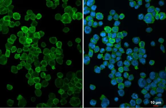
FCER1G antibody [N2C3] detects FCER1G protein at cytoplasm by immunofluorescent analysis. Sample: THP-1 cells were fixed in 4% paraformaldehyde at RT for 15 min. Green: FCER1G stained by FCER1G antibody [N2C3] (GTX108487) diluted at 1:500. Blue: Fluoroshield with DAPI (GTX30920).
![Non-transfected (–) and transfected (+) 293T whole cell extracts (30 μg) were separated by 15% SDS-PAGE, and the membrane was blotted with FCER1G antibody [N2C3] (GTX108487) diluted at 1:5000. The HRP-conjugated anti-rabbit IgG antibody (GTX213110-01) was used to detect the primary antibody.](https://www.genetex.com/upload/website/prouct_img/normal/GTX108487/GTX108487_44496_20220325_WB_B_w_23060120_111.webp "Non-transfected (–) and transfected (+) 293T whole cell extracts (30 μg) were separated by 15% SDS-PAGE, and the membrane was blotted with FCER1G antibody [N2C3] (GTX108487) diluted at 1:5000. The HRP-conjugated anti-rabbit IgG antibody (GTX213110-01) was used to detect the primary antibody.")
![FCER1G antibody [N2C3] detects FCER1G protein at cell membrane by immunofluorescent analysis. Sample: HepG2 cells were fixed in 4% paraformaldehyde at RT for 15 min. Green: FCER1G stained by FCER1G antibody [N2C3] (GTX108487) diluted at 1:500. Red: alpha Tubulin, a cytoskeleton marker, stained by alpha Tubulin antibody [GT114] (GTX628802) diluted at 1:1000. Blue: Fluoroshield with DAPI (GTX30920). Scale bar= 10μm.](https://www.genetex.com/upload/website/prouct_img/normal/GTX108487/GTX108487_44538_20220321_ICC_IF_w_23060120_931.webp "FCER1G antibody [N2C3] detects FCER1G protein at cell membrane by immunofluorescent analysis. Sample: HepG2 cells were fixed in 4% paraformaldehyde at RT for 15 min. Green: FCER1G stained by FCER1G antibody [N2C3] (GTX108487) diluted at 1:500. Red: alpha Tubulin, a cytoskeleton marker, stained by alpha Tubulin antibody [GT114] (GTX628802) diluted at 1:1000. Blue: Fluoroshield with DAPI (GTX30920). Scale bar= 10μm.")
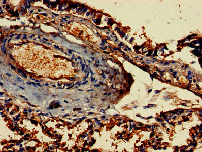
 antibody at 1:100 dilution.
Antigen Retrieval: Trilogy? (EDTA based, pH 8.0) buffer, 15min")
![Various whole cell extracts (30 μg) were separated by 15% SDS-PAGE, and the membrane was blotted with FCER1G antibody [N2C3] (GTX108487) diluted at 1:1000. The HRP-conjugated anti-rabbit IgG antibody (GTX213110-01) was used to detect the primary antibody.](https://www.genetex.com/upload/website/prouct_img/normal/GTX108487/GTX108487_44496_20211203_WB_H_M_w_23060120_598.webp "Various whole cell extracts (30 μg) were separated by 15% SDS-PAGE, and the membrane was blotted with FCER1G antibody [N2C3] (GTX108487) diluted at 1:1000. The HRP-conjugated anti-rabbit IgG antibody (GTX213110-01) was used to detect the primary antibody.")
![Various whole cell extracts (30 μg) were separated by 15% SDS-PAGE, and the membrane was blotted with FCER1G antibody [N2C3] (GTX108487) diluted at 1:2000. The HRP-conjugated anti-rabbit IgG antibody (GTX213110-01) was used to detect the primary antibody. Corresponding RNA expression data for the same cell lines are based on Human Protein Atlas program.](https://www.genetex.com/upload/website/prouct_img/normal/GTX108487/GTX108487_44496_20230602_WB_TPM_watermark_23060622_299.webp "Various whole cell extracts (30 μg) were separated by 15% SDS-PAGE, and the membrane was blotted with FCER1G antibody [N2C3] (GTX108487) diluted at 1:2000. The HRP-conjugated anti-rabbit IgG antibody (GTX213110-01) was used to detect the primary antibody. Corresponding RNA expression data for the same cell lines are based on Human Protein Atlas program.")
FCER1G antibody [N2C3] detects FCER1G protein at cytoplasm by immunofluorescent analysis. Sample: THP-1 cells were fixed in 4% paraformaldehyde at RT for 15 min. Green: FCER1G stained by FCER1G antibody [N2C3] (GTX108487) diluted at 1:500. Blue: Fluoroshield with DAPI (GTX30920).
FCER1G antibody [N2C3]
GTX108487
ApplicationsImmunoFluorescence, ImmunoPrecipitation, Western Blot, ImmunoCytoChemistry, ImmunoHistoChemistry, ImmunoHistoChemistry Paraffin
Product group Antibodies
ReactivityHuman, Mouse
TargetFCER1G
Overview
- SupplierGeneTex
- Product NameFCER1G antibody [N2C3]
- Delivery Days Customer9
- Application Supplier NoteWB: 1:500-1:3000. IHC-P: 1:100-1:1000. *Optimal dilutions/concentrations should be determined by the researcher.Not tested in other applications.
- ApplicationsImmunoFluorescence, ImmunoPrecipitation, Western Blot, ImmunoCytoChemistry, ImmunoHistoChemistry, ImmunoHistoChemistry Paraffin
- CertificationResearch Use Only
- ClonalityPolyclonal
- Concentration0.73 mg/ml
- ConjugateUnconjugated
- Gene ID2207
- Target nameFCER1G
- Target descriptionFc epsilon receptor Ig
- Target synonymsFCRG, high affinity immunoglobulin epsilon receptor subunit gamma, Fc fragment of IgE receptor Ig, Fc fragment of IgE, high affinity I, receptor for; gamma polypeptide, Fc receptor gamma-chain, FcepsilonRI gamma chain, FcepsilonRIgamma, fc-epsilon RI-gamma, fcRgamma, fceRI gamma, immunoglobulin E receptor, high affinity, gamma chain
- HostRabbit
- IsotypeIgG
- Protein IDP30273
- Protein NameHigh affinity immunoglobulin epsilon receptor subunit gamma
- Scientific DescriptionThe high affinity IgE receptor is a key molecule involved in allergic reactions. It is a tetramer composed of 1 alpha, 1 beta, and 2 gamma chains. The gamma chains are also subunits of other Fc receptors. [provided by RefSeq]
- ReactivityHuman, Mouse
- Storage Instruction-20°C or -80°C,2°C to 8°C
- UNSPSC41116161
Datasheet
Related products
Product group Antibodies
Anti-FCER1G Antibody144-60112
ApplicationsWestern Blot
ReactivityHuman, Mouse, Rat
TargetFCER1G
- SizePrice
Product group Antibodies

FCER1G AntibodyABX031280
ApplicationsWestern Blot, ELISA, ImmunoHistoChemistry
- SizePrice
Product group Antibodies

Anti-Fc epsilon R [IB10]AB01396-1.1-BT
ApplicationsFlow Cytometry, Neutralisation/Blocking
ReactivityHuman
TargetFCER1G
- SizePrice
Product group Antibodies

FCER1G AntibodyLS-C747959
ApplicationsWestern Blot
ReactivityHuman, Mouse, Rat
TargetFCER1G
- SizePrice
Product group Antibodies
FCER1G AntibodyCSB-PA008533LA01HU
ApplicationsImmunoFluorescence, ELISA, ImmunoHistoChemistry
ReactivityHuman
TargetFCER1G
- SizePrice
Product group Antibodies
Fcer1G Polyclonal AntibodyCAC08430
ApplicationsImmunoFluorescence, ELISA, ImmunoHistoChemistry
TargetFCER1G
- SizePrice
Product group Antibodies
Anti-FCER1G AntibodyHPA026872
ApplicationsImmunoHistoChemistry
ReactivityHuman
TargetFCER1G
- SizePrice
![FCER1G antibody [HL1418] detects FCER1G protein by immunohistochemical analysis. Sample: Paraffin-embedded rat tissues. FCER1G stained by FCER1G antibody [HL1418] (GTX636884) diluted at 1:100. Antigen Retrieval: Citrate buffer, pH 6.0, 15 min](https://www.genetex.com/upload/website/prouct_img/normal/GTX636884/GTX636884_44683_20221227_IHC-P_multiple_R_22122821_683.webp)
Product group Antibodies
FCER1G antibody [HL1418]GTX636884
ApplicationsImmunoFluorescence, ImmunoPrecipitation, Western Blot, ImmunoCytoChemistry, ImmunoHistoChemistry, ImmunoHistoChemistry Paraffin
ReactivityCanine, Feline, Human, Mouse, Rat
TargetFCER1G
- SizePrice