

Ferritin Heavy Chain antibody [N1C3] detects Ferritin Heavy Chain protein at cytoplasm by immunohistochemical analysis. Sample: Paraffin-embedded mouse liver. Ferritin Heavy Chain stained by Ferritin Heavy Chain antibody [N1C3] (GTX101733) diluted at 1:500. Antigen Retrieval: Citrate buffer, pH 6.0, 15 min
![Ferritin Heavy Chain antibody [N1C3] detects Ferritin Heavy Chain protein at cytoplasm by immunohistochemical analysis. Sample: Paraffin-embedded rat brain. Ferritin Heavy Chain stained by Ferritin Heavy Chain antibody [N1C3] (GTX101733) diluted at 1:500. Antigen Retrieval: Citrate buffer, pH 6.0, 15 min](https://www.genetex.com/upload/website/prouct_img/normal/GTX101733/GTX101733_44699_20220701_IHC-P_R_22071401_116.webp "Ferritin Heavy Chain antibody [N1C3] detects Ferritin Heavy Chain protein at cytoplasm by immunohistochemical analysis. Sample: Paraffin-embedded rat brain. Ferritin Heavy Chain stained by Ferritin Heavy Chain antibody [N1C3] (GTX101733) diluted at 1:500. Antigen Retrieval: Citrate buffer, pH 6.0, 15 min")
![Various whole cell extracts (30 μg) were separated by 12% SDS-PAGE, and the membrane was blotted with Ferritin Heavy Chain antibody [N1C3] (GTX101733) diluted at 1:3000. The HRP-conjugated anti-rabbit IgG antibody (GTX213110-01) was used to detect the primary antibody, and the signal was developed with Trident ECL plus-Enhanced. Corresponding RNA expression data for the same cell lines are based on Human Protein Atlas program.](https://www.genetex.com/upload/website/prouct_img/normal/GTX101733/GTX101733_44482_20221111_WB_TPM_watermark_22110919_553.webp "Various whole cell extracts (30 μg) were separated by 12% SDS-PAGE, and the membrane was blotted with Ferritin Heavy Chain antibody [N1C3] (GTX101733) diluted at 1:3000. The HRP-conjugated anti-rabbit IgG antibody (GTX213110-01) was used to detect the primary antibody, and the signal was developed with Trident ECL plus-Enhanced. Corresponding RNA expression data for the same cell lines are based on Human Protein Atlas program.")
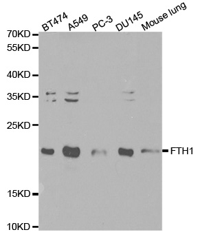
![Various tissue extracts (50 μg) were separated by 12% SDS-PAGE, and the membrane was blotted with Ferritin Heavy Chain antibody [N1C3] (GTX101733) diluted at 1:20000. The HRP-conjugated anti-rabbit IgG antibody (GTX213110-01) was used to detect the primary antibody.](https://www.genetex.com/upload/website/prouct_img/normal/GTX101733/GTX101733_44216_20211217_WB_M_R_w_23060100_202.webp "Various tissue extracts (50 μg) were separated by 12% SDS-PAGE, and the membrane was blotted with Ferritin Heavy Chain antibody [N1C3] (GTX101733) diluted at 1:20000. The HRP-conjugated anti-rabbit IgG antibody (GTX213110-01) was used to detect the primary antibody.")
![Human tissue extract (30 μg) was separated by 12% SDS-PAGE, and the membrane was blotted with Ferritin Heavy Chain antibody [N1C3] (GTX101733) diluted at 1:500.](https://www.genetex.com/upload/website/prouct_img/normal/GTX101733/GTX101733_42529_20160728_WB_H_liver_w_23060100_222.webp "Human tissue extract (30 μg) was separated by 12% SDS-PAGE, and the membrane was blotted with Ferritin Heavy Chain antibody [N1C3] (GTX101733) diluted at 1:500.")
![Ferritin Heavy Chain antibody [N1C3] detects Ferritin Heavy Chain protein at cytoplasm and nucleus by immunofluorescent analysis. Sample: HeLa cells were fixed in 4% paraformaldehyde at RT for 15 min. Green: Ferritin Heavy Chain protein stained by Ferritin Heavy Chain antibody [N1C3] (GTX101733) diluted at 1:500. Red: Phalloidin, a cytoskeleton marker, diluted at 1:50. Blue: Hoechst 33342 staining. Scale bar = 10 μm.](https://www.genetex.com/upload/website/prouct_img/normal/GTX101733/GTX101733_42592_20161123_IFA_w_23060100_930.webp "Ferritin Heavy Chain antibody [N1C3] detects Ferritin Heavy Chain protein at cytoplasm and nucleus by immunofluorescent analysis. Sample: HeLa cells were fixed in 4% paraformaldehyde at RT for 15 min. Green: Ferritin Heavy Chain protein stained by Ferritin Heavy Chain antibody [N1C3] (GTX101733) diluted at 1:500. Red: Phalloidin, a cytoskeleton marker, diluted at 1:50. Blue: Hoechst 33342 staining. Scale bar = 10 μm.")
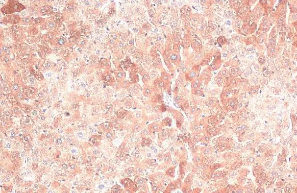
![Ferritin Heavy Chain antibody [N1C3] detects Ferritin Heavy Chain protein at cytoplasm in rat liver by immunohistochemical analysis. Sample: Paraffin-embedded rat liver. Ferritin Heavy Chain antibody [N1C3] (GTX101733) diluted at 1:500.
Antigen Retrieval: Citrate buffer, pH 6.0, 15 min](https://www.genetex.com/upload/website/prouct_img/normal/GTX101733/GTX101733_42529_20160823_IHC-P_R_w_23060100_922.webp "Ferritin Heavy Chain antibody [N1C3] detects Ferritin Heavy Chain protein at cytoplasm in rat liver by immunohistochemical analysis. Sample: Paraffin-embedded rat liver. Ferritin Heavy Chain antibody [N1C3] (GTX101733) diluted at 1:500.
Antigen Retrieval: Citrate buffer, pH 6.0, 15 min")
![Rat tissue extract (50 μg) was separated by 12% SDS-PAGE, and the membrane was blotted with Ferritin Heavy Chain antibody [N1C3] (GTX101733) diluted at 1:500. The HRP-conjugated anti-rabbit IgG antibody (GTX213110-01) was used to detect the primary antibody.](https://www.genetex.com/upload/website/prouct_img/normal/GTX101733/GTX101733_44734_20220708_WB_R_liver_25012200_733.webp "Rat tissue extract (50 μg) was separated by 12% SDS-PAGE, and the membrane was blotted with Ferritin Heavy Chain antibody [N1C3] (GTX101733) diluted at 1:500. The HRP-conjugated anti-rabbit IgG antibody (GTX213110-01) was used to detect the primary antibody.")
![Various whole cell extracts (30 μg) were separated by 12% SDS-PAGE, and the membrane was blotted with Ferritin Heavy Chain antibody [N1C3] (GTX101733) diluted at 1:1000. The HRP-conjugated anti-rabbit IgG antibody (GTX213110-01) was used to detect the primary antibody.](https://www.genetex.com/upload/website/prouct_img/normal/GTX101733/GTX101733_44734_20220708_WB_25012200_670.webp "Various whole cell extracts (30 μg) were separated by 12% SDS-PAGE, and the membrane was blotted with Ferritin Heavy Chain antibody [N1C3] (GTX101733) diluted at 1:1000. The HRP-conjugated anti-rabbit IgG antibody (GTX213110-01) was used to detect the primary antibody.")
Ferritin Heavy Chain antibody [N1C3] detects Ferritin Heavy Chain protein at cytoplasm by immunohistochemical analysis. Sample: Paraffin-embedded mouse liver. Ferritin Heavy Chain stained by Ferritin Heavy Chain antibody [N1C3] (GTX101733) diluted at 1:500. Antigen Retrieval: Citrate buffer, pH 6.0, 15 min
Ferritin Heavy Chain antibody [N1C3]
GTX101733
ApplicationsImmunoFluorescence, Western Blot, ImmunoCytoChemistry, ImmunoHistoChemistry, ImmunoHistoChemistry Paraffin
Product group Antibodies
ReactivityHuman, Mouse, Rat
TargetFTH1
Overview
- SupplierGeneTex
- Product NameFerritin Heavy Chain antibody [N1C3]
- Delivery Days Customer9
- Application Supplier NoteWB: 1:500-1:3000. ICC/IF: 1:100-1:1000. IHC-P: 1:100-1:1000. *Optimal dilutions/concentrations should be determined by the researcher.Not tested in other applications.
- ApplicationsImmunoFluorescence, Western Blot, ImmunoCytoChemistry, ImmunoHistoChemistry, ImmunoHistoChemistry Paraffin
- CertificationResearch Use Only
- ClonalityPolyclonal
- Concentration1.22 mg/ml
- ConjugateUnconjugated
- Gene ID2495
- Target nameFTH1
- Target descriptionferritin heavy chain 1
- Target synonymsFHC, FTH, FTHL6, HFE5, NBIA9, PIG15, PLIF, ferritin heavy chain, H-ferritin, apoferritin, cell proliferation-inducing gene 15 protein, ferritin H subunit, ferritin, heavy polypeptide 1, placenta immunoregulatory factor, proliferation-inducing protein 15
- HostRabbit
- IsotypeIgG
- Protein IDP02794
- Protein NameFerritin heavy chain
- Scientific DescriptionThis gene encodes the heavy subunit of ferritin, the major intracellular iron storage protein in prokaryotes and eukaryotes. It is composed of 24 subunits of the heavy and light ferritin chains. Variation in ferritin subunit composition may affect the rates of iron uptake and release in different tissues. A major function of ferritin is the storage of iron in a soluble and nontoxic state. Defects in ferritin proteins are associated with several neurodegenerative diseases. This gene has multiple pseudogenes. Several alternatively spliced transcript variants have been observed, but their biological validity has not been determined. [provided by RefSeq]
- ReactivityHuman, Mouse, Rat
- Storage Instruction-20°C or -80°C,2°C to 8°C
- UNSPSC41116161
Datasheet
Related products
Product group Antibodies
Anti-FTH1 AntibodyA29425
ApplicationsImmunoFluorescence, Western Blot, ImmunoHistoChemistry
ReactivityHuman, Mouse, Rat
- SizePrice
Product group Antibodies
Anti-Ferritin (N-term) Antibody130-10047
ApplicationsELISA
ReactivityHuman
TargetFTH1
- SizePrice
Product group Antibodies

Anti-Ferritin H [H107]AB03941-1.1
ApplicationsImmunoPrecipitation, ELISA
ReactivityHuman
TargetFTH1
- SizePrice
Product group Antibodies
References
ApplicationsFlow Cytometry, Western Blot, ELISA, ImmunoHistoChemistry, ImmunoHistoChemistry Frozen, ImmunoHistoChemistry Paraffin
ReactivityBovine, Canine, Equine, Human, Mouse, Porcine, Rabbit, Rat, Sheep
TargetFTH1
- SizePrice
Product group Antibodies
FTH1 AntibodyCSB-PA008485
ApplicationsWestern Blot, ELISA
ReactivityHuman, Mouse, Rat
TargetFTH1
- SizePrice
Product group Antibodies

Goat anti-FTH1EB09091
ApplicationsWestern Blot, ELISA, ImmunoHistoChemistry
ReactivityHuman
TargetFTH1
- SizePrice
Product group Antibodies
ApplicationsImmunoFluorescence, Western Blot, ImmunoCytoChemistry, ImmunoHistoChemistry
ReactivityMouse
TargetFTH1
- SizePrice

Product group Antibodies
References
ApplicationsWestern Blot, ImmunoHistoChemistry
ReactivityHuman
TargetFTH1
- SizePrice
Product group Antibodies

ApplicationsELISA
ReactivityHuman
TargetFTH1
- SizePrice