

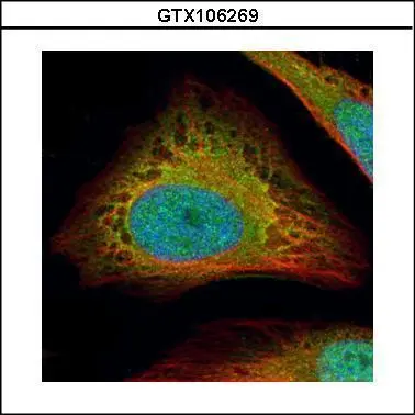
Confocal immunofluorescence analysis (Olympus FV10i) of paraformaldehyde-fixed HeLa, using Flightless I(GTX106269) antibody (Green) at 1:500 dilution. Alpha-tubulin filaments were labeled with GTX11304 (Red) at 1:2000.
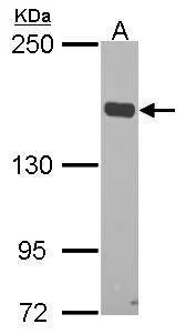
 A:Raji (GTX27908) 5% SDS PAGE GTX106269 diluted at 1:1000")
Confocal immunofluorescence analysis (Olympus FV10i) of paraformaldehyde-fixed HeLa, using Flightless I(GTX106269) antibody (Green) at 1:500 dilution. Alpha-tubulin filaments were labeled with GTX11304 (Red) at 1:2000.
Flightless I antibody [N3C1], Internal
GTX106269
ApplicationsImmunoFluorescence, Western Blot, ImmunoCytoChemistry
Product group Antibodies
ReactivityHuman
TargetFLII
Overview
- SupplierGeneTex
- Product NameFlightless I antibody [N3C1], Internal
- Delivery Days Customer9
- Application Supplier NoteWB: 1:500-1:3000. ICC/IF: 1:100-1:1000. *Optimal dilutions/concentrations should be determined by the researcher.Not tested in other applications.
- ApplicationsImmunoFluorescence, Western Blot, ImmunoCytoChemistry
- CertificationResearch Use Only
- ClonalityPolyclonal
- Concentration0.88 mg/ml
- ConjugateUnconjugated
- Gene ID2314
- Target nameFLII
- Target descriptionFLII actin remodeling protein
- Target synonymsCMD2J, FLI, FLIL, Fli1, protein flightless-1 homolog, flightless I actin binding protein, flightless I homolog
- HostRabbit
- IsotypeIgG
- Protein IDQ13045
- Protein NameProtein flightless-1 homolog
- Scientific DescriptionThis gene encodes a protein with a gelsolin-like actin binding domain and an N-terminal leucine-rich repeat-protein protein interaction domain. The protein is similar to a Drosophila protein involved in early embryogenesis and the structural organization of indirect flight muscle. The gene is located within the Smith-Magenis syndrome region on chromosome 17. [provided by RefSeq]
- ReactivityHuman
- Storage Instruction-20°C or -80°C,2°C to 8°C
- UNSPSC41116161
Datasheet
Related products
Product group Antibodies
FLII Monoclonal AntibodyBSM-60437M
ApplicationsImmunoFluorescence, Western Blot, ImmunoCytoChemistry, ImmunoHistoChemistry, ImmunoHistoChemistry Frozen, ImmunoHistoChemistry Paraffin
ReactivityHuman, Mouse, Rat
TargetFLII
- SizePrice
Product group Antibodies
FLII AntibodyCSB-PA615661LA01HU
ApplicationsImmunoFluorescence, ELISA, ImmunoHistoChemistry
ReactivityHuman
TargetFLII
- SizePrice

Product group Antibodies
ApplicationsWestern Blot, ImmunoHistoChemistry
ReactivityHuman, Mouse, Rat
TargetFLII
- SizePrice
Product group Antibodies

FLII / FLI AntibodyLS-C750244
ApplicationsWestern Blot
ReactivityHuman
TargetFLII
- SizePrice

Product group Antibodies
Anti-FLII AntibodyHPA007084
ApplicationsWestern Blot, ImmunoCytoChemistry, ImmunoHistoChemistry
ReactivityHuman, Mouse, Rat
TargetFLII
- SizePrice
Product group Antibodies
ApplicationsWestern Blot, ELISA, ImmunoCytoChemistry, ImmunoHistoChemistry, ImmunoHistoChemistry Frozen, ImmunoHistoChemistry Paraffin
ReactivityMouse, Porcine, Rat
TargetFLII
- SizePrice
![Flightless I antibody [HL2965] detects Flightless I protein by immunofluorescent analysis. Sample: HeLa cells were fixed in ice-cold MeOH for 5 min. Green: Flightless I stained by Flightless I antibody [HL2965] (GTX640352) diluted at 1:500. Red: alpha Tubulin, a cytoskeleton marker, stained by alpha Tubulin antibody [GT114] (GTX628802) diluted at 1:1000. Blue: Fluoroshield with DAPI (GTX30920).](https://www.genetex.com/upload/website/prouct_img/normal/GTX640352/GTX640352_T-45411_20240607_ICC_IF_24062501_993.webp)
Product group Antibodies
Flightless I antibody [HL2965]GTX640352
ApplicationsImmunoFluorescence, Western Blot, ImmunoCytoChemistry
ReactivityHuman
TargetFLII
- SizePrice
![Flightless I antibody [HL2966] detects Flightless I protein by immunofluorescent analysis. Sample: HeLa cells were fixed in ice-cold MeOH for 5 min. Green: Flightless I stained by Flightless I antibody [HL2966] (GTX640353) diluted at 1:500. Red: alpha Tubulin, a cytoskeleton marker, stained by alpha Tubulin antibody [GT114] (GTX628802) diluted at 1:1000. Blue: Fluoroshield with DAPI (GTX30920).](https://www.genetex.com/upload/website/prouct_img/normal/GTX640353/GTX640353_T-45411_20240607_ICC_IF_24062501_689.webp)
Product group Antibodies
Flightless I antibody [HL2966]GTX640353
ApplicationsImmunoFluorescence, Western Blot, ImmunoCytoChemistry, ImmunoHistoChemistry, ImmunoHistoChemistry Paraffin
ReactivityHuman, Mouse, Rat
TargetFLII
- SizePrice
![Whole tissue extract (50 μg) was separated by 5% SDS-PAGE, and the membrane was blotted with Flightless I antibod [HL3438] (GTX641311) diluted at 1:1000. The HRP-conjugated anti-rabbit IgG antibody (GTX213110-01) was used to detect the primary antibody.](https://www.genetex.com/upload/website/prouct_img/normal/GTX641311/GTX641311_T-45593_20241115_WB_M_muscle_24111918_945.webp)
Product group Antibodies
Flightless I antibody [HL3438]GTX641311
ApplicationsImmunoFluorescence, Western Blot, ImmunoCytoChemistry
ReactivityHuman, Mouse
TargetFLII
- SizePrice
Product group Antibodies
Flightless I antibody [C2C3], C-termGTX114410
ApplicationsImmunoFluorescence, Western Blot, ImmunoCytoChemistry
ReactivityHuman, Mouse, Rat
TargetFLII
- SizePrice