

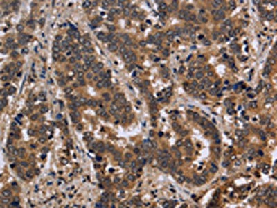
The image on the left is immunohistochemistry of paraffin-embedded Human liver cancer tissue using CSB-PA916967(FMR1 Antibody) at dilution 1/30, on the right is treated with fusion protein. (Original magnification: x200)
The image on the left is immunohistochemistry of paraffin-embedded Human liver cancer tissue using CSB-PA916967(FMR1 Antibody) at dilution 1/30, on the right is treated with fusion protein. (Original magnification: x200)
FMR1 Antibody
CSB-PA916967
ApplicationsELISA, ImmunoHistoChemistry
Product group Antibodies
ReactivityHuman, Mouse, Rat
TargetFMR1
Overview
- SupplierCusabio
- Product NameFMR1 Antibody
- Delivery Days Customer20
- ApplicationsELISA, ImmunoHistoChemistry
- CertificationResearch Use Only
- ClonalityPolyclonal
- ConjugateUnconjugated
- Gene ID2332
- Target nameFMR1
- Target descriptionfragile X messenger ribonucleoprotein 1
- Target synonymsFMRP, FRAXA, POF, POF1, fragile X messenger ribonucleoprotein 1, FMRP translational regulator 1, synaptic functional regulator FMR1
- HostRabbit
- IsotypeIgG
- Protein IDQ06787
- Protein NameFragile X messenger ribonucleoprotein 1
- Scientific DescriptionThe protein encoded by this gene binds RNA and is associated with polysomes. The encoded protein may be involved in mRNA trafficking from the nucleus to the cytoplasm. A trinucleotide repeat (CGG) in the 5 UTR is normally found at 6-53 copies, but an expansion to 55-230 repeats is the cause of fragile X syndrome. Expansion of the trinucleotide repeat may also cause one form of premature ovarian failure (POF1). Multiple alternatively spliced transcript variants that encode different protein isoforms and which are located in different cellular locations have been described for this gene.
- ReactivityHuman, Mouse, Rat
- Storage Instruction-20°C or -80°C
- UNSPSC41116161
Related products
Product group Antibodies
Anti-FMR1 AntibodyA30850
ApplicationsImmunoFluorescence, Western Blot, ImmunoHistoChemistry
ReactivityHuman, Mouse, Rat
- SizePrice
Product group Antibodies
Anti-FMR1 Antibody144-06092
ApplicationsWestern Blot, ImmunoHistoChemistry
ReactivityHuman, Mouse, Rat
TargetFMR1
- SizePrice
Product group Antibodies

FMR1 / FMRP AntibodyLS-C746851
ApplicationsWestern Blot
ReactivityHuman, Rat
TargetFMR1
- SizePrice
Product group Antibodies
FMR1 Recombinant Antibody, AbBy Fluor-488 ConjugatedBSM-61466R-BF488
ApplicationsFlow Cytometry, ImmunoFluorescence, Western Blot
ReactivityHuman, Mouse, Rat
TargetFMR1
- SizePrice
Product group Antibodies

Goat anti-FMR1EB07868
ApplicationsWestern Blot, ELISA, ImmunoHistoChemistry
ReactivityCanine, Human, Mouse, Rat
TargetFMR1
- SizePrice
Product group Antibodies
ApplicationsImmunoPrecipitation, Western Blot, ImmunoCytoChemistry, ImmunoHistoChemistry
ReactivityMouse, Rat
TargetFMR1
- SizePrice
Product group Antibodies
FMRP antibodyGTX134734
ApplicationsWestern Blot
ReactivityHuman
TargetFMR1
- SizePrice

Product group Antibodies
Anti-FMR1 AntibodyHPA050118
ApplicationsImmunoCytoChemistry, ImmunoHistoChemistry
ReactivityHuman
TargetFMR1
- SizePrice
Product group Antibodies
ApplicationsWestern Blot, ELISA, ImmunoHistoChemistry, ImmunoHistoChemistry Paraffin
ReactivityHuman
TargetFMR1
- SizePrice