

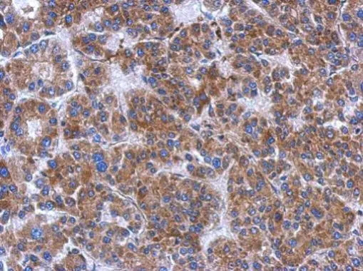
Immunohistochemical analysis of paraffin-embedded human hepatoma, using FN3K(GTX107579) antibody at 1:500 dilution.
Antigen Retrieval: Trilogy? (EDTA based, pH 8.0) buffer, 15min
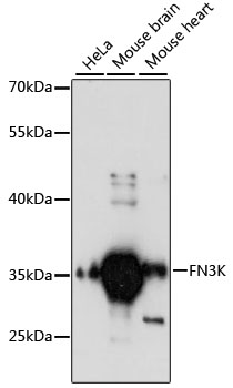
![Various whole cell extracts (30 μg) were separated by 10% SDS-PAGE, and the membrane was blotted with FN3K antibody [N1C3-2] (GTX107579) diluted at 1:1000. The HRP-conjugated anti-rabbit IgG antibody (GTX213110-01) was used to detect the primary antibody.](https://www.genetex.com/upload/website/prouct_img/normal/GTX107579/GTX107579_44615_20220318_WB_25041720_456.webp "Various whole cell extracts (30 μg) were separated by 10% SDS-PAGE, and the membrane was blotted with FN3K antibody [N1C3-2] (GTX107579) diluted at 1:1000. The HRP-conjugated anti-rabbit IgG antibody (GTX213110-01) was used to detect the primary antibody.")
![Various whole cell extracts (30 μg) were separated by 10% SDS-PAGE, and the membrane was blotted with FN3K antibody [N1C3-2] (GTX107579) diluted at 1:1000. The HRP-conjugated anti-rabbit IgG antibody (GTX213110-01) was used to detect the primary antibody. Corresponding RNA expression data for the same cell lines are based on Human Protein Atlas program.](https://www.genetex.com/upload/website/prouct_img/normal/GTX107579/GTX107579_44615_20230224_WB_TPM_watermark_25041720_179.webp "Various whole cell extracts (30 μg) were separated by 10% SDS-PAGE, and the membrane was blotted with FN3K antibody [N1C3-2] (GTX107579) diluted at 1:1000. The HRP-conjugated anti-rabbit IgG antibody (GTX213110-01) was used to detect the primary antibody. Corresponding RNA expression data for the same cell lines are based on Human Protein Atlas program.")
Immunohistochemical analysis of paraffin-embedded human hepatoma, using FN3K(GTX107579) antibody at 1:500 dilution.
Antigen Retrieval: Trilogy? (EDTA based, pH 8.0) buffer, 15min
FN3K antibody [N1C3-2]
GTX107579
ApplicationsWestern Blot, ImmunoHistoChemistry, ImmunoHistoChemistry Paraffin
Product group Antibodies
ReactivityHuman
TargetFN3K
Overview
- SupplierGeneTex
- Product NameFN3K antibody [N1C3-2]
- Delivery Days Customer9
- Application Supplier NoteWB: 1:500-1:3000. IHC-P: 1:100-1:1000. *Optimal dilutions/concentrations should be determined by the researcher.Not tested in other applications.
- ApplicationsWestern Blot, ImmunoHistoChemistry, ImmunoHistoChemistry Paraffin
- CertificationResearch Use Only
- ClonalityPolyclonal
- Concentration0.35 mg/ml
- ConjugateUnconjugated
- Gene ID64122
- Target nameFN3K
- Target descriptionfructosamine 3 kinase
- Target synonymsfructosamine-3-kinase, protein-psicosamine 3-kinase FN3K, protein-ribulosamine 3-kinase FN3K
- HostRabbit
- IsotypeIgG
- Protein IDQ9H479
- Protein NameFructosamine-3-kinase
- Scientific DescriptionFN3K catalyzes phosphorylation of fructosamines formed by glycation, the nonenzymatic reaction of glucose with primary amines followed by Amadori rearrangement. Phosphorylation of fructosamines may initiate metabolism of the modified amine and result in deglycation of glycated proteins (Delpierre et al., 2000 [PubMed 11016445]).[supplied by OMIM]
- ReactivityHuman
- Storage Instruction-20°C or -80°C,2°C to 8°C
- UNSPSC41116161
Datasheet
Related products
Product group Antibodies
FN3K AntibodyCSB-PA008760GA01HU
ApplicationsWestern Blot, ELISA
ReactivityHuman
TargetFN3K
- SizePrice
Product group Antibodies
ApplicationsWestern Blot, ELISA
ReactivityHuman, Mouse
TargetFN3K
- SizePrice
Product group Antibodies
Anti-FN3K AntibodyA89361
ApplicationsWestern Blot
ReactivityHuman, Mouse
- SizePrice
Product group Antibodies

FN3K / Fructosamine-3-Kinase AntibodyLS-C748760
ApplicationsWestern Blot
ReactivityHuman, Mouse
TargetFN3K
- SizePrice

Product group Antibodies
Anti-FN3K AntibodyHPA059151
ApplicationsImmunoCytoChemistry
ReactivityHuman
TargetFN3K
- SizePrice
Product group Antibodies
ApplicationsImmunoPrecipitation, Western Blot, ImmunoCytoChemistry, ImmunoHistoChemistry
ReactivityMouse, Porcine, Rat
TargetFN3K
- SizePrice
![FN3K antibody [HL2223] detects FN3K protein at cytoplasm by immunohistochemical analysis. Sample: Paraffin-embedded mouse stomach. FN3K stained by FN3K antibody [HL2223] (GTX638268) diluted at 1:200. Antigen Retrieval: Citrate buffer, pH 6.0, 15 min](https://www.genetex.com/upload/website/prouct_img/normal/GTX638268/GTX638268_T-44956_20230324_IHC-P_M_23032819_105.webp)
Product group Antibodies
FN3K antibody [HL2223]GTX638268
ApplicationsWestern Blot, ImmunoHistoChemistry, ImmunoHistoChemistry Paraffin
ReactivityHuman, Mouse, Rat
TargetFN3K
- SizePrice
Product group Antibodies
FN3K Polyclonal AntibodyBS-13189R
ApplicationsImmunoFluorescence, Western Blot, ELISA, ImmunoCytoChemistry, ImmunoHistoChemistry, ImmunoHistoChemistry Frozen, ImmunoHistoChemistry Paraffin
ReactivityHuman, Mouse, Rat
TargetFN3K
- SizePrice