

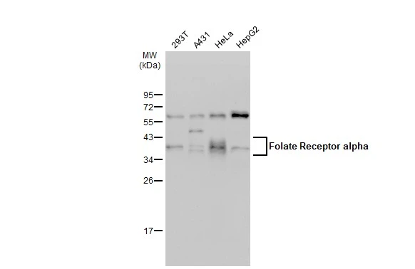
Various whole cell extracts (30 μg) were separated by 12% SDS-PAGE, and the membrane was blotted with Folate Receptor alpha antibody (GTX134660) diluted at 1:500. The HRP-conjugated anti-rabbit IgG antibody (GTX213110-01) was used to detect the primary antibody, and the signal was developed with Trident ECL plus-Enhanced.
![Folate Receptor alpha antibody detects Folate Receptor alpha protein at cytoplasm by immunofluorescent analysis. Sample: HeLa cells were fixed in 4% paraformaldehyde at RT for 15 min. Green: Folate Receptor alpha stained by Folate Receptor alpha antibody (GTX134660) diluted at 1:500. Red: alpha Tubulin, a cytoskeleton marker, stained by alpha Tubulin antibody [GT114] (GTX628802) diluted at 1:1000. Blue: Fluoroshield with DAPI (GTX30920). Scale bar= 10μm.](https://www.genetex.com/upload/website/prouct_img/normal/GTX134660/GTX134660_43481_20220321_ICC_IF_w_23060620_321.webp "Folate Receptor alpha antibody detects Folate Receptor alpha protein at cytoplasm by immunofluorescent analysis. Sample: HeLa cells were fixed in 4% paraformaldehyde at RT for 15 min. Green: Folate Receptor alpha stained by Folate Receptor alpha antibody (GTX134660) diluted at 1:500. Red: alpha Tubulin, a cytoskeleton marker, stained by alpha Tubulin antibody [GT114] (GTX628802) diluted at 1:1000. Blue: Fluoroshield with DAPI (GTX30920). Scale bar= 10μm.")
 and transfected (+) 293T whole cell extracts (30 μg) were separated by 12% SDS-PAGE, and the membrane was blotted with Folate Receptor alpha antibody (GTX134660) diluted at 1:1000. The HRP-conjugated anti-rabbit IgG antibody (GTX213110-01) was used to detect the primary antibody.")
 were separated by 12% SDS-PAGE, and the membrane was blotted with Folate Receptor alpha antibody (GTX134660) diluted at 1:500. The HRP-conjugated anti-rabbit IgG antibody (GTX213110-01) was used to detect the primary antibody. Corresponding RNA expression data for the same cell lines are based on Human Protein Atlas program.")
Various whole cell extracts (30 μg) were separated by 12% SDS-PAGE, and the membrane was blotted with Folate Receptor alpha antibody (GTX134660) diluted at 1:500. The HRP-conjugated anti-rabbit IgG antibody (GTX213110-01) was used to detect the primary antibody, and the signal was developed with Trident ECL plus-Enhanced.
Folate Receptor alpha antibody
GTX134660
ApplicationsImmunoFluorescence, Western Blot, ImmunoCytoChemistry
Product group Antibodies
ReactivityHuman
TargetFOLR1
Overview
- SupplierGeneTex
- Product NameFolate Receptor alpha antibody
- Delivery Days Customer9
- Application Supplier NoteWB: 1:500-1:3000. *Optimal dilutions/concentrations should be determined by the researcher.Not tested in other applications.
- ApplicationsImmunoFluorescence, Western Blot, ImmunoCytoChemistry
- CertificationResearch Use Only
- ClonalityPolyclonal
- Concentration0.64 mg/ml
- ConjugateUnconjugated
- Gene ID2348
- Target nameFOLR1
- Target descriptionfolate receptor alpha
- Target synonymsFBP, FOLR, FRalpha, NCFTD, folate receptor alpha, FR-alpha, KB cells FBP, adult folate-binding protein, folate binding protein, folate receptor 1 (adult), folate receptor, adult, ovarian tumor-associated antigen MOv18
- HostRabbit
- IsotypeIgG
- Protein IDP15328
- Protein NameFolate receptor alpha
- Scientific DescriptionThe protein encoded by this gene is a member of the folate receptor family. Members of this gene family bind folic acid and its reduced derivatives, and transport 5-methyltetrahydrofolate into cells. This gene product is a secreted protein that either anchors to membranes via a glycosyl-phosphatidylinositol linkage or exists in a soluble form. Mutations in this gene have been associated with neurodegeneration due to cerebral folate transport deficiency. Due to the presence of two promoters, multiple transcription start sites, and alternative splicing, multiple transcript variants encoding the same protein have been found for this gene. [provided by RefSeq, Oct 2009]
- ReactivityHuman
- Storage Instruction-20°C or -80°C,2°C to 8°C
- UNSPSC41116161
Datasheet
Related products
Product group Antibodies
Anti-FOLR1 AntibodyA97535
ApplicationsWestern Blot, ELISA
ReactivityHuman, Mouse, Rat
- SizePrice
Product group Antibodies

Anti-FOLR1 [Farletuzumab (MORAb-003; M3)]Ab03043-10.0
ApplicationsFunctional Assay, ELISA, ImmunoHistoChemistry, Neutralisation/Blocking, Other Application
ReactivityHuman, Monkey
TargetFOLR1
- SizePrice
Product group Antibodies
ApplicationsFlow Cytometry
TargetFOLR1
- SizePrice
Product group Antibodies

FOLR1 / Folate Receptor Alpha AntibodyLS-C831294
ApplicationsELISA, ImmunoHistoChemistry
ReactivityHuman, Mouse
TargetFOLR1
- SizePrice
Product group Antibodies
ApplicationsWestern Blot
ReactivityHuman, Mouse, Rat
TargetFOLR1
- SizePrice
Product group Antibodies
FOLR1 AntibodyCSB-PA008378
ApplicationsWestern Blot, ELISA
ReactivityHuman, Mouse, Rat
TargetFOLR1
- SizePrice
Product group Antibodies
ApplicationsWestern Blot, ImmunoHistoChemistry
ReactivityMouse
TargetFOLR1
- SizePrice

Product group Antibodies
References
ApplicationsWestern Blot
ReactivityHuman, Mouse, Rat
TargetFOLR1
- SizePrice