

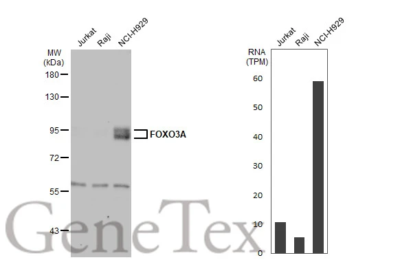
Various whole cell extracts (30 μg) were separated by 7.5% SDS-PAGE, and the membrane was blotted with FOXO3A antibody [C3], C-term (GTX100277) diluted at 1:1000. The HRP-conjugated anti-rabbit IgG antibody (GTX213110-01) was used to detect the primary antibody. Corresponding RNA expression data for the same cell lines are based on Human Protein Atlas program.
![Non-transfected (–) and transfected (+) C2C12 whole cell extracts (30 μg) were separated by 7.5% SDS-PAGE, and the membrane was blotted with FOXO3A antibody [C3], C-term (GTX100277) diluted at 1:500. The HRP-conjugated anti-rabbit IgG antibody (GTX213110-01) was used to detect the primary antibody.](https://www.genetex.com/upload/website/prouct_img/normal/GTX100277/GTX100277_39443_20160303_WB_M_shRNA_watermark_w_23060100_531.webp "Non-transfected (–) and transfected (+) C2C12 whole cell extracts (30 μg) were separated by 7.5% SDS-PAGE, and the membrane was blotted with FOXO3A antibody [C3], C-term (GTX100277) diluted at 1:500. The HRP-conjugated anti-rabbit IgG antibody (GTX213110-01) was used to detect the primary antibody.")
![FOXO3A antibody [C3], C-term detects FOXO3A protein at nucleus by immunofluorescent analysis. Sample: HeLa cells were fixed in 4% paraformaldehyde at RT for 15 min. Green: FOXO3A stained by FOXO3A antibody [C3], C-term (GTX100277) diluted at 1:1000. Blue: Hoechst 33342 staining. Scale bar= 10 μm.](https://www.genetex.com/upload/website/prouct_img/normal/GTX100277/GTX100277_43489_20190703_ICC_IF_w_23060100_586.webp "FOXO3A antibody [C3], C-term detects FOXO3A protein at nucleus by immunofluorescent analysis. Sample: HeLa cells were fixed in 4% paraformaldehyde at RT for 15 min. Green: FOXO3A stained by FOXO3A antibody [C3], C-term (GTX100277) diluted at 1:1000. Blue: Hoechst 33342 staining. Scale bar= 10 μm.")
![FOXO3A antibody [C3], C-term detects FOXO3A protein at cytoplasm and nucleus by immunohistochemical analysis. Sample: Paraffin-embedded mouse brain. FOXO3A stained by FOXO3A antibody [C3], C-term (GTX100277) diluted at 1:2000. Antigen Retrieval: Citrate buffer, pH 6.0, 15 min](https://www.genetex.com/upload/website/prouct_img/normal/GTX100277/GTX100277_43292_20200327_IHC-P_M_w_23060100_842.webp "FOXO3A antibody [C3], C-term detects FOXO3A protein at cytoplasm and nucleus by immunohistochemical analysis. Sample: Paraffin-embedded mouse brain. FOXO3A stained by FOXO3A antibody [C3], C-term (GTX100277) diluted at 1:2000. Antigen Retrieval: Citrate buffer, pH 6.0, 15 min")
![FOXO3A antibody [C3], C-term detects FOXO3A protein at cytoplasm and nucleus by immunohistochemical analysis. Sample: Paraffin-embedded mouse duodenum. FOXO3A stained by FOXO3A antibody [C3], C-term (GTX100277) diluted at 1:2000. Antigen Retrieval: Citrate buffer, pH 6.0, 15 min](https://www.genetex.com/upload/website/prouct_img/normal/GTX100277/GTX100277_43489_20200327_IHC-P_M_w_23060100_125.webp "FOXO3A antibody [C3], C-term detects FOXO3A protein at cytoplasm and nucleus by immunohistochemical analysis. Sample: Paraffin-embedded mouse duodenum. FOXO3A stained by FOXO3A antibody [C3], C-term (GTX100277) diluted at 1:2000. Antigen Retrieval: Citrate buffer, pH 6.0, 15 min")
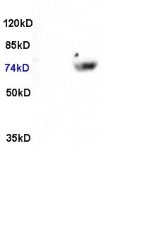
 A: Mouse brain 7.5% SDS PAGE GTX100277 diluted at 1:500 The HRP-conjugated anti-rabbit IgG antibody (GTX213110-01) was used to detect the primary antibody.")
![FOXO3A antibody [C3], C-term detects FOXO3A protein at nucleus by immunohistochemical analysis. Sample: Paraffin-embedded mouse testis. FOXO3A stained by FOXO3A antibody [C3], C-term (GTX100277) diluted at 1:500. Antigen Retrieval: Citrate buffer, pH 6.0, 15 min](https://www.genetex.com/upload/website/prouct_img/normal/GTX100277/GTX100277_43985_20200828_IHC-P_M_w_23060100_144.webp "FOXO3A antibody [C3], C-term detects FOXO3A protein at nucleus by immunohistochemical analysis. Sample: Paraffin-embedded mouse testis. FOXO3A stained by FOXO3A antibody [C3], C-term (GTX100277) diluted at 1:500. Antigen Retrieval: Citrate buffer, pH 6.0, 15 min")
 antibody at 1:500 dilution.
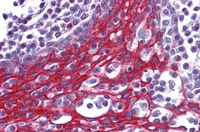
Antigen Retrieval: Trilogy? (EDTA based, pH 8.0) buffer, 15min")
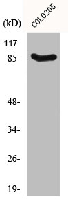
 5% SDS-PAGE The immunoprecipitated FOXO3A protein was detected by FOXO3A antibody (GTX101150) diluted at 1:1000. EasyBlot anti-rabbit IgG (GTX221666-01) was used as a secondary reagent.")
![Various whole cell extracts (30 μg) were separated by 7.5% SDS-PAGE, and the membrane was blotted with FOXO3A antibody [C3], C-term (GTX100277) diluted at 1:1000. The HRP-conjugated anti-rabbit IgG antibody (GTX213110-01) was used to detect the primary antibody.](https://www.genetex.com/upload/website/prouct_img/normal/GTX100277/GTX100277_43489_20220107_WB_w_23060100_988.webp "Various whole cell extracts (30 μg) were separated by 7.5% SDS-PAGE, and the membrane was blotted with FOXO3A antibody [C3], C-term (GTX100277) diluted at 1:1000. The HRP-conjugated anti-rabbit IgG antibody (GTX213110-01) was used to detect the primary antibody.")
Various whole cell extracts (30 μg) were separated by 7.5% SDS-PAGE, and the membrane was blotted with FOXO3A antibody [C3], C-term (GTX100277) diluted at 1:1000. The HRP-conjugated anti-rabbit IgG antibody (GTX213110-01) was used to detect the primary antibody. Corresponding RNA expression data for the same cell lines are based on Human Protein Atlas program.
FOXO3A antibody [C3], C-term
GTX100277
ApplicationsImmunoFluorescence, ImmunoPrecipitation, Western Blot, ImmunoCytoChemistry, ImmunoHistoChemistry, ImmunoHistoChemistry Paraffin
Product group Antibodies
ReactivityHuman, Mammals, Mouse, Rat, Xenopus
TargetFOXO3
Overview
- SupplierGeneTex
- Product NameFOXO3A antibody [C3], C-term
- Delivery Days Customer9
- Application Supplier NoteWB: 1:500-1:3000. ICC/IF: 1:100-1:1000. IHC-P: 1:100-1:1000. IP: 1:100-1:500. *Optimal dilutions/concentrations should be determined by the researcher.Not tested in other applications.
- ApplicationsImmunoFluorescence, ImmunoPrecipitation, Western Blot, ImmunoCytoChemistry, ImmunoHistoChemistry, ImmunoHistoChemistry Paraffin
- CertificationResearch Use Only
- ClonalityPolyclonal
- Concentration0.56 mg/ml
- ConjugateUnconjugated
- Gene ID2309
- Target nameFOXO3
- Target descriptionforkhead box O3
- Target synonymsAF6q21, FKHRL1, FKHRL1P2, FOXO2, FOXO3A, forkhead box protein O3, FOXO3A-, forkhead box O3A, forkhead homolog (rhabdomyosarcoma) like 1, forkhead in rhabdomyosarcoma-like 1, forkhead, Drosophila, homolog of, in rhabdomyosarcoma-like 1
- HostRabbit
- IsotypeIgG
- Protein IDO43524
- Protein NameForkhead box protein O3
- Scientific DescriptionThis gene belongs to the forkhead family of transcription factors which are characterized by a distinct forkhead domain. This gene likely functions as a trigger for apoptosis through expression of genes necessary for cell death. Translocation of this gene with the MLL gene is associated with secondary acute leukemia. Alternatively spliced transcript variants encoding the same protein have been observed. [provided by RefSeq]
- ReactivityHuman, Mammals, Mouse, Rat, Xenopus
- Storage Instruction-20°C or -80°C,2°C to 8°C
- UNSPSC41116161
Datasheet
Related products

Product group Antibodies
Anti-FOXO3A AntibodyA98640
ApplicationsELISA, ImmunoHistoChemistry
ReactivityHuman, Mouse
- SizePrice
Product group Antibodies
Anti-Phospho-FOXO3-S253 Antibody144-50758
ApplicationsWestern Blot
ReactivityHuman, Mouse, Rat
TargetFOXO3
- SizePrice

Product group Antibodies
Anti-FOXO3 AntibodyAMAB91872
ApplicationsWestern Blot, ImmunoHistoChemistry
ReactivityHuman
TargetFOXO3
- SizePrice
Product group Antibodies

FOXO3 / FOXO3A Antibody (Ser253)LS-C769243
ApplicationsImmunoFluorescence, Western Blot, ELISA, ImmunoHistoChemistry, ImmunoHistoChemistry Paraffin
ReactivityHuman, Mouse, Rat
TargetFOXO3
- SizePrice

Product group Antibodies
Anti-FOXO3A/FOXO3 Antibody Picoband(r)A00252-2-CARRIER-FREE
ApplicationsWestern Blot, ELISA
ReactivityHuman
TargetFOXO3
- SizePrice
Product group Antibodies
References
FOXO3A Polyclonal AntibodyBS-1548R
ApplicationsImmunoFluorescence, Western Blot, ELISA, ImmunoHistoChemistry, ImmunoHistoChemistry Frozen, ImmunoHistoChemistry Paraffin
ReactivityHuman, Mouse, Rat
TargetFOXO3
- SizePrice
Product group Antibodies
FOXO3 AntibodyCSB-PA002574
ApplicationsImmunoFluorescence, Western Blot, ELISA, ImmunoHistoChemistry
ReactivityHuman, Mouse, Rat
TargetFOXO3
- SizePrice
Product group Antibodies

ApplicationsWestern Blot, ELISA, ImmunoHistoChemistry
ReactivityBovine, Canine, Human, Mouse, Porcine, Rat
TargetFOXO3
- SizePrice
Product group Antibodies
ApplicationsImmunoPrecipitation, Western Blot, ImmunoCytoChemistry, ImmunoHistoChemistry
ReactivityMouse, Porcine, Rat
TargetFOXO3
- SizePrice
Product group Antibodies
FOXO3A antibody, C-termGTX17026
ApplicationsWestern Blot, ImmunoHistoChemistry, ImmunoHistoChemistry Paraffin
ReactivityHuman
TargetFOXO3
- SizePrice