

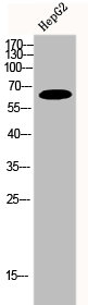
Whole cell extract (30 μg) was separated by 10% SDS-PAGE, and the membrane was blotted with FRS2 antibody [HL1774] (GTX637426) diluted at 1:5000. The HRP-conjugated anti-rabbit IgG antibody (GTX213110-01) was used to detect the primary antibody.
![Various tissue extracts (50 μg) were separated by 10% SDS-PAGE, and the membrane was blotted with FRS2 antibody [HL1774] (GTX637426) diluted at 1:5000. The HRP-conjugated anti-rabbit IgG antibody (GTX213110-01) was used to detect the primary antibody.](https://www.genetex.com/upload/website/prouct_img/normal/GTX637426/GTX637426_T-44802_20220930_WB_M_tissue_22101319_561.webp "Various tissue extracts (50 μg) were separated by 10% SDS-PAGE, and the membrane was blotted with FRS2 antibody [HL1774] (GTX637426) diluted at 1:5000. The HRP-conjugated anti-rabbit IgG antibody (GTX213110-01) was used to detect the primary antibody.")
![Various whole cell extracts (30 μg) were separated by 10% SDS-PAGE, and the membrane was blotted with FRS2 antibody [HL1774] (GTX637426) diluted at 1:1000. The HRP-conjugated anti-rabbit IgG antibody (GTX213110-01) was used to detect the primary antibody.](https://www.genetex.com/upload/website/prouct_img/normal/GTX637426/GTX637426_T-44802_20221014_WB_R_22101718_836.webp "Various whole cell extracts (30 μg) were separated by 10% SDS-PAGE, and the membrane was blotted with FRS2 antibody [HL1774] (GTX637426) diluted at 1:1000. The HRP-conjugated anti-rabbit IgG antibody (GTX213110-01) was used to detect the primary antibody.")
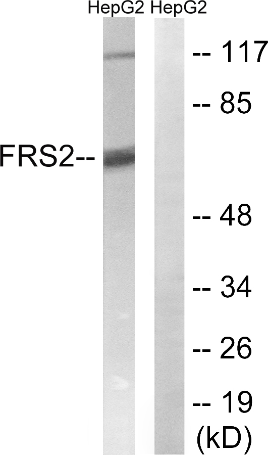
![Non-transfected (–) and transfected (+) 293T whole cell extracts (30 μg) were separated by 10% SDS-PAGE, and the membrane was blotted with FRS2 antibody [HL1774] (GTX637426) diluted at 1:50000. The HRP-conjugated anti-rabbit IgG antibody (GTX213110-01) was used to detect the primary antibody.](https://www.genetex.com/upload/website/prouct_img/normal/GTX637426/GTX637426_T-44802_20221125_WB_shRNA_watermark_22112723_168.webp "Non-transfected (–) and transfected (+) 293T whole cell extracts (30 μg) were separated by 10% SDS-PAGE, and the membrane was blotted with FRS2 antibody [HL1774] (GTX637426) diluted at 1:50000. The HRP-conjugated anti-rabbit IgG antibody (GTX213110-01) was used to detect the primary antibody.")
![Various whole cell extracts (30 μg) were separated by 10% SDS-PAGE, and the membrane was blotted with FRS2 antibody [HL1774] (GTX637426) diluted at 1:5000. The HRP-conjugated anti-rabbit IgG antibody (GTX213110-01) was used to detect the primary antibody.](https://www.genetex.com/upload/website/prouct_img/normal/GTX637426/GTX637426_44865_20221125_WB_H_M_22112723_197.webp "Various whole cell extracts (30 μg) were separated by 10% SDS-PAGE, and the membrane was blotted with FRS2 antibody [HL1774] (GTX637426) diluted at 1:5000. The HRP-conjugated anti-rabbit IgG antibody (GTX213110-01) was used to detect the primary antibody.")
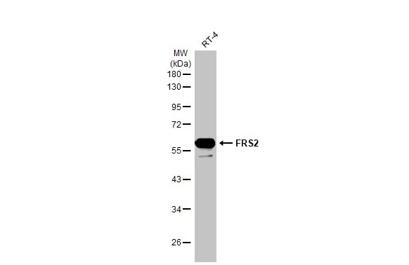
Whole cell extract (30 μg) was separated by 10% SDS-PAGE, and the membrane was blotted with FRS2 antibody [HL1774] (GTX637426) diluted at 1:5000. The HRP-conjugated anti-rabbit IgG antibody (GTX213110-01) was used to detect the primary antibody.
FRS2 antibody [HL1774]
GTX637426
ApplicationsWestern Blot
Product group Antibodies
ReactivityHuman, Mouse, Rat
TargetFRS2
Overview
- SupplierGeneTex
- Product NameFRS2 antibody [HL1774]
- Delivery Days Customer9
- Application Supplier NoteWB: 1:1000-1:10000. *Optimal dilutions/concentrations should be determined by the researcher.Not tested in other applications.
- ApplicationsWestern Blot
- CertificationResearch Use Only
- ClonalityMonoclonal
- Clone IDHL1774
- Concentration1 mg/ml
- ConjugateUnconjugated
- Gene ID10818
- Target nameFRS2
- Target descriptionfibroblast growth factor receptor substrate 2
- Target synonymsFRS1A, FRS2A, FRS2alpha, SNT, SNT-1, SNT1, fibroblast growth factor receptor substrate 2, FGFR signalling adaptor, FGFR substrate 2, FGFR-signaling adaptor SNT, epididymis secretory sperm binding protein, suc1-associated neurotrophic factor target 1
- HostRabbit
- IsotypeIgG
- Protein IDQ8WU20
- Protein NameFibroblast growth factor receptor substrate 2
- ReactivityHuman, Mouse, Rat
- Storage Instruction-20°C or -80°C,2°C to 8°C
- UNSPSC41116161
Datasheet
Related products
Product group Antibodies
Anti-FRS2 AntibodyA99414
ApplicationsWestern Blot, ELISA
ReactivityHuman, Mouse
- SizePrice
Product group Antibodies

FRS2 (Phospho-Tyr436) AntibodyABX012649
ApplicationsWestern Blot, ELISA, ImmunoHistoChemistry
- SizePrice
Product group Antibodies
FRS2 Recombinant Antibody, Biotin ConjugatedBSM-61526R-BIOTIN
ApplicationsImmunoPrecipitation, Western Blot
ReactivityHuman
TargetFRS2
- SizePrice
Product group Antibodies
FRS2 AntibodyCSB-PA002584
ApplicationsWestern Blot, ELISA
ReactivityHuman, Mouse
TargetFRS2
- SizePrice
Product group Antibodies

Goat anti-FRS2EB12615
ApplicationsWestern Blot, ELISA
ReactivityBovine, Canine, Human, Mouse, Porcine
TargetFRS2
- SizePrice
Product group Antibodies
ApplicationsImmunoPrecipitation, Western Blot, ImmunoCytoChemistry, ImmunoHistoChemistry
ReactivityMouse, Rat
TargetFRS2
- SizePrice

Product group Antibodies
ApplicationsFlow Cytometry, ImmunoPrecipitation, Western Blot
ReactivityHuman
TargetFRS2
- SizePrice
Product group Antibodies

FRS2 Antibody (Internal)LS-C368751
ApplicationsWestern Blot
ReactivityHuman, Mouse
TargetFRS2
- SizePrice
Product group Antibodies
FRS2 antibodyGTX10425
ApplicationsImmunoPrecipitation, Western Blot
ReactivityHuman, Mouse, Rat
TargetFRS2
- SizePrice
Product group Antibodies
FRS2 (phospho Tyr436) antibodyGTX55340
ApplicationsWestern Blot, ImmunoHistoChemistry, ImmunoHistoChemistry Paraffin
ReactivityHuman
TargetFRS2
- SizePrice