

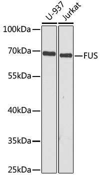
Various whole cell extracts (30 μg) were separated by 10% SDS-PAGE, and the membrane was blotted with FUS antibody [HL2454] (GTX638772) diluted at 1:1000. The HRP-conjugated anti-rabbit IgG antibody (GTX213110-01) was used to detect the primary antibody.
![FUS antibody [HL2454] detects FUS protein by immunohistochemical analysis. Sample: Paraffin-embedded rat tissues. FUS stained by FUS antibody [HL2454] (GTX638772) diluted at 1:100. Antigen Retrieval: Citrate buffer, pH 6.0, 15 min](https://www.genetex.com/upload/website/prouct_img/normal/GTX638772/GTX638772_T-45089_20230721_IHC-P_multiple_R_23073119_904.webp "FUS antibody [HL2454] detects FUS protein by immunohistochemical analysis. Sample: Paraffin-embedded rat tissues. FUS stained by FUS antibody [HL2454] (GTX638772) diluted at 1:100. Antigen Retrieval: Citrate buffer, pH 6.0, 15 min")
![FUS antibody [HL2454] detects FUS protein by immunohistochemical analysis. Sample: Paraffin-embedded mouse tissues. FUS stained by FUS antibody [HL2454] (GTX638772) diluted at 1:100. Antigen Retrieval: Citrate buffer, pH 6.0, 15 min](https://www.genetex.com/upload/website/prouct_img/normal/GTX638772/GTX638772_T-45089_20230721_IHC-P_multiple_M_23073119_734.webp "FUS antibody [HL2454] detects FUS protein by immunohistochemical analysis. Sample: Paraffin-embedded mouse tissues. FUS stained by FUS antibody [HL2454] (GTX638772) diluted at 1:100. Antigen Retrieval: Citrate buffer, pH 6.0, 15 min")
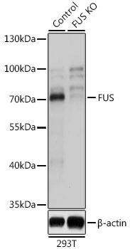
![Wild-type (WT) and FUS knockout (KO) 293T cell extracts (30 μg) were separated by 7.5% SDS-PAGE, and the membrane was blotted with FUS antibody [HL2454] (GTX638772) diluted at 1:3000. The HRP-conjugated anti-rabbit IgG antibody (GTX213110-01) was used to detect the primary antibody.](https://www.genetex.com/upload/website/prouct_img/normal/GTX638772/GTX638772_T-45089_20230728_WB_KO_watermark_23073119_464.webp "Wild-type (WT) and FUS knockout (KO) 293T cell extracts (30 μg) were separated by 7.5% SDS-PAGE, and the membrane was blotted with FUS antibody [HL2454] (GTX638772) diluted at 1:3000. The HRP-conjugated anti-rabbit IgG antibody (GTX213110-01) was used to detect the primary antibody.")
![Various whole cell extracts (30 μg) were separated by 10% SDS-PAGE, and the membrane was blotted with FUS antibody [HL2454] (GTX638772) diluted at 1:1000. The HRP-conjugated anti-rabbit IgG antibody (GTX213110-01) was used to detect the primary antibody.](https://www.genetex.com/upload/website/prouct_img/normal/GTX638772/GTX638772_45159_20230908_WB_23091901_439.webp "Various whole cell extracts (30 μg) were separated by 10% SDS-PAGE, and the membrane was blotted with FUS antibody [HL2454] (GTX638772) diluted at 1:1000. The HRP-conjugated anti-rabbit IgG antibody (GTX213110-01) was used to detect the primary antibody.")
![FUS antibody [HL2454] detects FUS protein at nucleus by immunofluorescent analysis. Sample: HepG2 cells were fixed in 4% PFA. Green: FUS stained by FUS antibody [HL2454] (GTX638772) diluted at 1:500. Blue: Fluoroshield with DAPI (GTX30920).](https://www.genetex.com/upload/website/prouct_img/normal/GTX638772/GTX638772_45159_20231229_ICC_IF_24011618_172.webp "FUS antibody [HL2454] detects FUS protein at nucleus by immunofluorescent analysis. Sample: HepG2 cells were fixed in 4% PFA. Green: FUS stained by FUS antibody [HL2454] (GTX638772) diluted at 1:500. Blue: Fluoroshield with DAPI (GTX30920).")
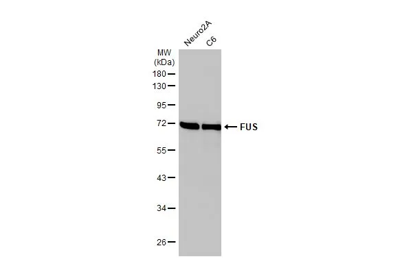
Various whole cell extracts (30 μg) were separated by 10% SDS-PAGE, and the membrane was blotted with FUS antibody [HL2454] (GTX638772) diluted at 1:1000. The HRP-conjugated anti-rabbit IgG antibody (GTX213110-01) was used to detect the primary antibody.
FUS antibody [HL2454]
GTX638772
ApplicationsImmunoFluorescence, Western Blot, ImmunoCytoChemistry, ImmunoHistoChemistry, ImmunoHistoChemistry Paraffin
Product group Antibodies
ReactivityHuman, Mouse, Rat
TargetFUS
Overview
- SupplierGeneTex
- Product NameFUS antibody [HL2454]
- Delivery Days Customer9
- Application Supplier NoteWB: 1:500-1:3000. *Optimal dilutions/concentrations should be determined by the researcher.Not tested in other applications.
- ApplicationsImmunoFluorescence, Western Blot, ImmunoCytoChemistry, ImmunoHistoChemistry, ImmunoHistoChemistry Paraffin
- CertificationResearch Use Only
- ClonalityMonoclonal
- Clone IDHL2454
- Concentration1 mg/ml
- ConjugateUnconjugated
- Gene ID2521
- Target nameFUS
- Target descriptionFUS RNA binding protein
- Target synonymsALS6, ETM4, FUS1, HNRNPP2, POMP75, TLS, altFUS, RNA-binding protein FUS, 75 kDa DNA-pairing protein, fus-like protein, fused in sarcoma, fusion gene in myxoid liposarcoma, heterogeneous nuclear ribonucleoprotein P2, oncogene FUS, oncogene TLS, translocated in liposarcoma protein
- HostRabbit
- IsotypeIgG
- Protein IDP35637
- Protein NameRNA-binding protein FUS
- Scientific DescriptionThis gene encodes a multifunctional protein component of the heterogeneous nuclear ribonucleoprotein (hnRNP) complex. The hnRNP complex is involved in pre-mRNA splicing and the export of fully processed mRNA to the cytoplasm. This protein belongs to the FET family of RNA-binding proteins which have been implicated in cellular processes that include regulation of gene expression, maintenance of genomic integrity and mRNA/microRNA processing. Alternative splicing results in multiple transcript variants. Defects in this gene result in amyotrophic lateral sclerosis type 6. [provided by RefSeq, Sep 2009]
- ReactivityHuman, Mouse, Rat
- Storage Instruction-20°C or -80°C,2°C to 8°C
- UNSPSC41116161
Datasheet
Related products
Product group Antibodies
Anti-TLS / FUS AntibodyA308899
ApplicationsImmunoPrecipitation, Western Blot, ImmunoHistoChemistry
ReactivityHuman, Mouse, Rat
- SizePrice

Product group Antibodies
Anti-TLS/FUS Antibody Picoband(r)A00771-1-CARRIER-FREE
ApplicationsWestern Blot
ReactivityHuman, Mouse, Rat
TargetFUS
- SizePrice
Product group Antibodies
Anti-FUS Antibody144-05921
ApplicationsWestern Blot, ImmunoHistoChemistry
ReactivityHuman, Mouse, Rat
TargetFUS
- SizePrice

Product group Antibodies
Anti-FUS AntibodyAMAB90549
ApplicationsWestern Blot, ImmunoCytoChemistry, ImmunoHistoChemistry
ReactivityHuman
TargetFUS
- SizePrice
Product group Antibodies

FUS / TLS AntibodyLS-C766686
ApplicationsELISA, ImmunoHistoChemistry
ReactivityHuman, Mouse
TargetFUS
- SizePrice
Product group Antibodies
TLS/FUS Recombinant Antibody, AbBy Fluor-350 ConjugatedBSM-61371R-BF350
ApplicationsImmunoFluorescence, Western Blot
ReactivityHuman, Mouse, Rat
TargetFUS
- SizePrice
Product group Antibodies
FUS Polyclonal AntibodyCAC14024
ApplicationsImmunoFluorescence, Western Blot, ELISA, ImmunoHistoChemistry
TargetFUS
- SizePrice
Product group Antibodies
FUS AntibodyCSB-PA02704A0RB
ApplicationsImmunoFluorescence, Western Blot, ELISA, ImmunoHistoChemistry
ReactivityHuman
TargetFUS
- SizePrice
Product group Antibodies
FUS antibodyGTX55629
ApplicationsImmunoFluorescence, ImmunoPrecipitation, Western Blot, ImmunoCytoChemistry, ImmunoHistoChemistry, ImmunoHistoChemistry Paraffin
ReactivityHuman, Mouse, Rat
TargetFUS
- SizePrice