

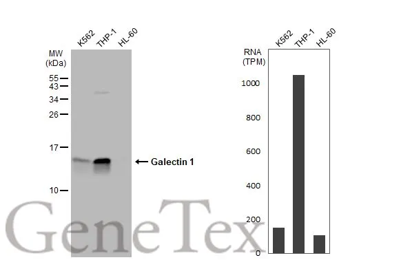
Various whole cell extracts (30 μg) were separated by 15% SDS-PAGE, and the membrane was blotted with Galectin 1 antibody (GTX116411) diluted at 1:2000. The HRP-conjugated anti-rabbit IgG antibody (GTX213110-01) was used to detect the primary antibody. Corresponding RNA expression data for the same cell lines are based on Human Protein Atlas program.
 A: NCI-H929 15% SDS PAGE GTX116411 diluted at 1:10000 The HRP-conjugated anti-rabbit IgG antibody (GTX213110-01) was used to detect the primary antibody.")
 antibody at 1:200 dilution.")
 dilution: 1:10000 The HRP-conjugated anti-rabbit IgG antibody (GTX213110-01) was used to detect the primary antibody.")

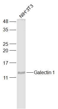
15% SDS-PAGE
The immunoprecipitated Galectin 1 protein was detected by Galectin 1 antibody (GTX116411) diluted at 1 : 1000.
EasyBlot anti-rabbit IgG (HRP) (GTX221666-01) was used as a secondary reagent.")
 A: BCL-1 15% SDS PAGE GTX116411 diluted at 1:10000 The HRP-conjugated anti-rabbit IgG antibody (GTX213110-01) was used to detect the primary antibody.")
 and Galectin 1 knockout (KO) 293T cell extracts (30 μg) were separated by 15% SDS-PAGE, and the membrane was blotted with Galectin 1 antibody (GTX116411) diluted at 1:500. The HRP-conjugated anti-rabbit IgG antibody (GTX213110-01) was used to detect the primary antibody, and the signal was developed with Trident ECL plus-Enhanced.")
 diluted at 1:200. Red: phalloidin, a cytoskeleton marker, stained by phalloidin (invitrogen, A12380) diluted at 1:200. Blue: Hoechst 33342 staining.")
 diluted at 1:500. Red: phalloidin, a cytoskeleton marker, stained by phalloidin (invitrogen, A12380) diluted at 1:200. Blue: Hoechst 33342 staining.")
Various whole cell extracts (30 μg) were separated by 15% SDS-PAGE, and the membrane was blotted with Galectin 1 antibody (GTX116411) diluted at 1:2000. The HRP-conjugated anti-rabbit IgG antibody (GTX213110-01) was used to detect the primary antibody. Corresponding RNA expression data for the same cell lines are based on Human Protein Atlas program.
Galectin 1 antibody
GTX116411
ApplicationsImmunoFluorescence, ImmunoPrecipitation, Western Blot, ImmunoCytoChemistry
Product group Antibodies
ReactivityHuman, Mouse, Rat
TargetLGALS1
Overview
- SupplierGeneTex
- Product NameGalectin 1 antibody
- Delivery Days Customer9
- Application Supplier NoteWB: 1:500-1:20000. ICC/IF: 1:100-1:1000. IP: 1:100-1:500. *Optimal dilutions/concentrations should be determined by the researcher.Not tested in other applications.
- ApplicationsImmunoFluorescence, ImmunoPrecipitation, Western Blot, ImmunoCytoChemistry
- CertificationResearch Use Only
- ClonalityPolyclonal
- Concentration1.48 mg/ml
- ConjugateUnconjugated
- Gene ID3956
- Target nameLGALS1
- Target descriptiongalectin 1
- Target synonymsGAL1, GBP, galectin-1, 14 kDa laminin-binding protein, 14 kDa lectin, HBL, HLBP14, HPL, S-Lac lectin 1, beta-galactoside-binding lectin L-14-I, beta-galactoside-binding protein 14kDa, epididymis secretory sperm binding protein, gal-1, galaptin, lactose-binding lectin 1, lectin, galactoside-binding, soluble, 1, putative MAPK-activating protein PM12
- HostRabbit
- IsotypeIgG
- Protein IDP09382
- Protein NameGalectin-1
- Scientific DescriptionThe galectins are a family of beta-galactoside-binding proteins implicated in modulating cell-cell and cell-matrix interactions. This gene product may act as an autocrine negative growth factor that regulates cell proliferation. [provided by RefSeq]
- ReactivityHuman, Mouse, Rat
- Storage Instruction-20°C or -80°C,2°C to 8°C
- UNSPSC12352203
References
- Paz H, Joo EJ, Chou CH, et al. Treatment of B-cell precursor acute lymphoblastic leukemia with the Galectin-1 inhibitor PTX008. J Exp Clin Cancer Res. 2018,37(1):67. doi: 10.1186/s13046-018-0721-7Read this paper
- Mori Y, Akita K, Yashiro M, et al. Binding of Galectin-3, a β-Galactoside-binding Lectin, to MUC1 Protein Enhances Phosphorylation of Extracellular Signal-regulated Kinase 1/2 (ERK1/2) and Akt, Promoting Tumor Cell Malignancy. J Biol Chem. 2015,290(43):26125-40. doi: 10.1074/jbc.M115.651489Read this paper
- May EW, Lin ST, Lin CC, et al. Identification of up- and down-regulated proteins in doxorubicin-resistant uterine cancer cells: reticulocalbin-1 plays a key role in the development of doxorubicin-associated resistance. Pharmacol Res. 2014,90:1-17. doi: 10.1016/j.phrs.2014.08.007Read this paper
- Lin LH, Chang SJ, Hu RY, et al. Biomarker discovery for neuroendocrine cervical cancer. Electrophoresis. 2014,35(14):2039-45. doi: 10.1002/elps.201400014Read this paper
- Fan CY, Chou HC, Lo YW, et al. Proteomic and redox-proteomic study on the role of glutathione reductase in human lung cancer cells. Electrophoresis. 2013,34(24):3305-14. doi: 10.1002/elps.201300250Read this paper
Datasheet
Related products
Product group Antibodies

Anti-LGALS1 [SAIC-25B-112]Ab00317-1.1
ApplicationsMass Spectrometry
ReactivityHuman
TargetLGALS1
- SizePrice

Product group Antibodies
Anti-LGALS1 Antibody144-01580
ApplicationsWestern Blot
ReactivityHuman, Mouse, Rat
TargetLGALS1
- SizePrice
![WB analysis of various samples using GTX02670 Galectin 1 antibody [GAL1/2499R]. Lane 1 : JEG-3 whole cell lysate Lane 2 : K562 whole cell lysate](https://www.genetex.com/upload/website/prouct_img/normal/GTX02670/GTX02670_20210319_WB_w_23053122_680.webp)
Product group Antibodies
Galectin 1 antibody [GAL1/2499R]GTX02670
ApplicationsWestern Blot, ELISA, ImmunoHistoChemistry, ImmunoHistoChemistry Paraffin, Other Application
ReactivityHuman
TargetLGALS1
- SizePrice
![Galectin 1 antibody detects Galectin 1 protein expression by immunohistochemical analysis. Sample: Frozen sectioned E13.5 Rat brain. Green: Galectin 1 protein stained by Galectin 1 antibody (GTX101566) diluted at 1:250. Red: beta Tubulin 3/ TUJ1, a mature neuron marker, stained by beta Tubulin 3/ TUJ1 antibody [GT11710] (GTX631836) diluted at 1:500. Blue: Fluoroshield with DAPI (GTX30920).](https://www.genetex.com/upload/website/prouct_img/normal/GTX101566/GTX101566_40744_20161005_IHC-Fr_R_w_23060100_981.webp)
Product group Antibodies
References
Galectin 1 antibodyGTX101566
ApplicationsImmunoFluorescence, ImmunoPrecipitation, Western Blot, ImmunoCytoChemistry, ImmunoHistoChemistry, ImmunoHistoChemistry Frozen, ImmunoHistoChemistry Paraffin
ReactivityHuman, Mouse, Rat
TargetLGALS1
- SizePrice
![Whole cell extract (30 μg) was separated by 15% SDS-PAGE, and the membrane was blotted with Galectin 1 antibody [GT2721] (GTX634454) diluted at 1:5000. The HRP-conjugated anti-mouse IgG antibody (GTX213111-01) was used to detect the primary antibody.](https://www.genetex.com/upload/website/prouct_img/normal/GTX634454/GTX634454_43094_20180119_WB_w_23061202_611.webp)
Product group Antibodies
Galectin 1 antibody [GT2721]GTX634454
ApplicationsWestern Blot
ReactivityHuman
TargetLGALS1
- SizePrice
Product group Antibodies
LGALS1 Polyclonal AntibodyCAC13100
ApplicationsImmunoFluorescence, Western Blot, ELISA, ImmunoHistoChemistry
ReactivityMouse
TargetLGALS1
- SizePrice
Product group Antibodies
References
Galectin 1 Polyclonal AntibodyBS-6594R
ApplicationsImmunoFluorescence, Western Blot, ELISA, ImmunoCytoChemistry, ImmunoHistoChemistry, ImmunoHistoChemistry Frozen, ImmunoHistoChemistry Paraffin
ReactivityBovine, Equine, Human, Mouse, Porcine, Rat, Sheep
TargetLGALS1
- SizePrice
Product group Antibodies
Anti-LGALS1 AntibodyA42202
ApplicationsWestern Blot
ReactivityHuman, Mouse, Rat
- SizePrice