

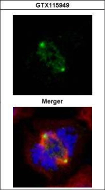
Confocal immunofluorescence analysis (Olympus FV10i) of paraformaldehyde-fixed 293T, using GCP4(GTX115949) antibody (Green) at 1:500 dilution. Alpha-tubulin filaments were labeled with GTX11304 (Red) at 1:500.
![GCP4 antibody detects GCP4 protein at cytoplasm and centrosome by immunofluorescent analysis. Sample: HeLa cells were fixed in 4% paraformaldehyde at RT for 15 min. Green: GCP4 protein stained by GCP4 antibody (GTX115949) diluted at 1:1000. Red: alpha Tubulin, a cytoskeleton marker, stained by alpha Tubulin antibody [GT114] (GTX628802) diluted at 1:1000. Blue: Hoechst 33342 staining.](https://www.genetex.com/upload/website/prouct_img/normal/GTX115949/GTX115949_40289_20150410_IFA_w_23060519_956.webp "GCP4 antibody detects GCP4 protein at cytoplasm and centrosome by immunofluorescent analysis. Sample: HeLa cells were fixed in 4% paraformaldehyde at RT for 15 min. Green: GCP4 protein stained by GCP4 antibody (GTX115949) diluted at 1:1000. Red: alpha Tubulin, a cytoskeleton marker, stained by alpha Tubulin antibody [GT114] (GTX628802) diluted at 1:1000. Blue: Hoechst 33342 staining.")
 dilution: 1:1000")
Confocal immunofluorescence analysis (Olympus FV10i) of paraformaldehyde-fixed 293T, using GCP4(GTX115949) antibody (Green) at 1:500 dilution. Alpha-tubulin filaments were labeled with GTX11304 (Red) at 1:500.
GCP4 antibody
GTX115949
ApplicationsImmunoFluorescence, Western Blot, ImmunoCytoChemistry, ImmunoHistoChemistry, ImmunoHistoChemistry Frozen
Product group Antibodies
ReactivityHuman, Mouse
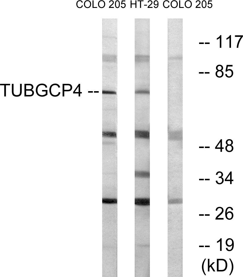
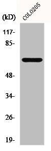
TargetTUBGCP4
Overview
- SupplierGeneTex
- Product NameGCP4 antibody
- Delivery Days Customer9
- Application Supplier NoteWB: 1:500-1:3000. ICC/IF: 1:100-1:1000. *Optimal dilutions/concentrations should be determined by the researcher.Not tested in other applications.
- ApplicationsImmunoFluorescence, Western Blot, ImmunoCytoChemistry, ImmunoHistoChemistry, ImmunoHistoChemistry Frozen
- CertificationResearch Use Only
- ClonalityPolyclonal
- Concentration0.75 mg/ml
- ConjugateUnconjugated
- Gene ID27229
- Target nameTUBGCP4
- Target descriptiontubulin gamma complex component 4
- Target synonyms76P, GCP-4, GCP4, Grip76, MCCRP3, gamma-tubulin complex component 4, gamma tubulin ring complex protein (76p gene), gamma-ring complex protein 76 kDa, gamma-tubulin complex protein 4, tubulin gamma complex associated protein 4
- HostRabbit
- IsotypeIgG
- Protein IDQ9UGJ1
- Protein NameGamma-tubulin complex component 4
- Scientific DescriptionGamma-tubulin complex is necessary for microtubule nucleation at the centrosome.
- ReactivityHuman, Mouse
- Storage Instruction-20°C or -80°C,2°C to 8°C
- UNSPSC12352203
References
- Li Z, Li H, Xu X, et al. Haploinsufficiency of GCP4 induces autophagy and leads to photoreceptor degeneration due to defective spindle assembly in retina. Cell Death Differ. 2020,27(2):556-572. doi: 10.1038/s41418-019-0371-0Read this paper
- Cota RR, Teixidó-Travesa N, Ezquerra A, et al. MZT1 regulates microtubule nucleation by linking γTuRC assembly to adapter-mediated targeting and activation. J Cell Sci. 2017,130(2):406-419. doi: 10.1242/jcs.195321Read this paper
- Lecland N, Lüders J. The dynamics of microtubule minus ends in the human mitotic spindle. Nat Cell Biol. 2014,16(8):770-8. doi: 10.1038/ncb2996Read this paper
Datasheet
Related products

Product group Antibodies
ApplicationsImmunoFluorescence, Western Blot, ELISA, ImmunoCytoChemistry, ImmunoHistoChemistry
TargetTUBGCP4
- SizePrice
![GCP4 antibody [GT812] detects GCP4 protein by Western blot analysis. A. 30 μg HeLa whole cell lysate/extract 10 % SDS-PAGE GCP4 antibody [GT812] (GTX629401) dilution: 1:1000](https://www.genetex.com/upload/website/prouct_img/normal/GTX629401/GTX629401_41323_WB_w_23061202_279.webp)
Product group Antibodies
GCP4 antibody [GT812]GTX629401
ApplicationsImmunoFluorescence, Western Blot, ImmunoCytoChemistry
ReactivityHuman
TargetTUBGCP4
- SizePrice
Product group Antibodies
TUBGCP4 Polyclonal AntibodyCAC13482
ApplicationsImmunoFluorescence, ImmunoPrecipitation, Western Blot, ELISA, ImmunoHistoChemistry
TargetTUBGCP4
- SizePrice
Product group Antibodies
GCP4 Polyclonal AntibodyBS-13320R
ApplicationsImmunoFluorescence, ELISA, ImmunoCytoChemistry, ImmunoHistoChemistry, ImmunoHistoChemistry Frozen, ImmunoHistoChemistry Paraffin
ReactivityBovine, Canine, Equine, Human, Mouse, Porcine, Rat
TargetTUBGCP4
- SizePrice
Product group Antibodies
Anti-TUBGCP4 AntibodyA98578
ApplicationsWestern Blot, ELISA
ReactivityHuman, Mouse
- SizePrice
Product group Antibodies
TUBGCP4 AntibodyCSB-PA002661
ApplicationsWestern Blot, ELISA, ImmunoHistoChemistry
ReactivityHuman, Mouse
TargetTUBGCP4
- SizePrice
Product group Antibodies

TUBGCP4 AntibodyLS-C832038
ApplicationsWestern Blot, ELISA
ReactivityHuman, Mouse
TargetTUBGCP4
- SizePrice

Product group Antibodies
Anti-TUBGCP4 AntibodyHPA043212
ApplicationsWestern Blot, ImmunoCytoChemistry, ImmunoHistoChemistry
ReactivityHuman
TargetTUBGCP4
- SizePrice