

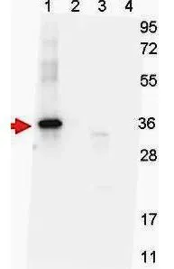
Western blot shows detection of recombinant NAG-1 protein present in Pichia pastoris whole cell lysates: lane 1 - yeast cell lysate expressing NAG-1 H variant with SUMO expression tag at 36 kDa; lane 2 - yeast cell lysate expressing NAG-1 D variant with SUMO expression tag at 36 kDa; lane 3 - yeast cell lysate expressing NAG-1 H variant; and lane 4 - yeast cell lysate expressing NAG-1 D variant. All lysates were run under reducing conditions. Primary antibody was used at a 1:1,000 dilution in TBS containing 1% BSA and 0.2% Tween, and reacted overnight at 4oC. For detection, a 1:40,000 dilution of peroxidase conjugated Goat-anti-Mouse IgG secondary antibody for 30 min at room temperature. Molecular weight estimation was made by comparison to prestained MW markers.
![ELISA analysis of various samples using serially diluted GTX48499 GDF15 (H Variant) antibody [7C12.B3.F2]. Red : H variant Green : D variant Coating : 0.1 μg](https://www.genetex.com/upload/website/prouct_img/normal/GTX48499/GTX48499_20240423_ELISA_243_24042320_788.webp "ELISA analysis of various samples using serially diluted GTX48499 GDF15 (H Variant) antibody [7C12.B3.F2]. Red : H variant Green : D variant Coating : 0.1 μg")
![WB analysis of various samples using GTX48499 GDF15 (H Variant) antibody [7C12.B3.F2]. Lane 1 : SUMO-tagged NAG-1 H variant Lane 2 : SUMO-tagged NAG-1 D variant Lane 3 : NAG-1 H variant Lane 4 : NAG-1 D variant Dilution : 1:1000](https://www.genetex.com/upload/website/prouct_img/normal/GTX48499/GTX48499_20240423_WB_244_24042320_379.webp "WB analysis of various samples using GTX48499 GDF15 (H Variant) antibody [7C12.B3.F2]. Lane 1 : SUMO-tagged NAG-1 H variant Lane 2 : SUMO-tagged NAG-1 D variant Lane 3 : NAG-1 H variant Lane 4 : NAG-1 D variant Dilution : 1:1000")
Western blot shows detection of recombinant NAG-1 protein present in Pichia pastoris whole cell lysates: lane 1 - yeast cell lysate expressing NAG-1 H variant with SUMO expression tag at 36 kDa; lane 2 - yeast cell lysate expressing NAG-1 D variant with SUMO expression tag at 36 kDa; lane 3 - yeast cell lysate expressing NAG-1 H variant; and lane 4 - yeast cell lysate expressing NAG-1 D variant. All lysates were run under reducing conditions. Primary antibody was used at a 1:1,000 dilution in TBS containing 1% BSA and 0.2% Tween, and reacted overnight at 4oC. For detection, a 1:40,000 dilution of peroxidase conjugated Goat-anti-Mouse IgG secondary antibody for 30 min at room temperature. Molecular weight estimation was made by comparison to prestained MW markers.
GDF15 (H Variant) antibody [7C12.B3.F2]
GTX48499
ApplicationsWestern Blot, ELISA
Product group Antibodies
ReactivityHuman
TargetGDF15
Overview
- SupplierGeneTex
- Product NameGDF15 (H Variant) antibody [7C12.B3.F2]
- Delivery Days Customer9
- Application Supplier NoteWB: 1:1000. ELISA: 1:150000. *Optimal dilutions/concentrations should be determined by the researcher.Not tested in other applications.
- ApplicationsWestern Blot, ELISA
- CertificationResearch Use Only
- ClonalityMonoclonal
- Clone ID7C12.B3.F2
- Concentration1.3 mg/ml
- ConjugateUnconjugated
- Gene ID9518
- Target nameGDF15
- Target descriptiongrowth differentiation factor 15
- Target synonymsGDF-15, HG, MIC-1, MIC1, NAG-1, PDF, PLAB, PTGFB, growth/differentiation factor 15, NRG-1, NSAID (nonsteroidal anti-inflammatory drug)-activated protein 1, NSAID-activated gene 1 protein, NSAID-regulated gene 1 protein, PTGF-beta, macrophage inhibitory cytokine 1, non-steroidal anti-inflammatory drug-activated gene-1, placental TGF-beta, placental bone morphogenetic protein, prostate differentiation factor
- HostMouse
- IsotypeIgG2b
- Protein IDQ99988
- Protein NameGrowth/differentiation factor 15
- Scientific DescriptionNon-steroidal anti-inflammatory drug (NSAID) activated gene (NAG-1) is a member of the transforming growth factor-beta (TGF-beta) superfamily. NAG-1 is also known as Macrophage Inhibitory Cytokine-1 (MIC-1), Growth Differentiation Factor 15 (GDF15), Placental Bone Morphogenetic Protein (PLAB), or Prostate Derived Factor (PDF). NAG-1 is expressed in human placenta, prostate and colon. It possesses antitumorigenic and proapoptotic activities. NAG-1 expression is dramatically increased in inflammation, injury and malignancy. Increase of NAG-1 expression is a feature of many cancers including breast, colon, pancreas and prostate. In a number of studies, NAG-1 expression was increased by a number of NSAIDs. This increase in expression may correlate with the chemopreventive effect NSAIDs seem to have with certain cancers. NAG-1 expression is also induced by PPAR gamma ligands and by several dietary compounds such as conjugated linoleic acids (CLAs), naturally occurring fatty acids in ruminant food products, indoles, epicatechin gallate, and genistein. Induced expression of NAG-1 results in stimulation of apoptosis and inhibition of cell growth. Inhibition of NAG-1 induced expression by small interference RNA (siRNA) results in repression of induced apoptosis. NAG-1 expression is regulated by a numbers of transcription factors such as ERG-1 and Sp1. EGR-1 may be necessary for NSAID-induced NAG-1 expression. The study of expression of NAG-1 proteins, including variants, is important to define their potential role as serum biomarkers for cancer diagnosis, treatment monitoring, epidemiology study, and nutrition surveys.
- ReactivityHuman
- Storage Instruction-20°C or -80°C,2°C to 8°C
- UNSPSC41116161
Datasheet
Related products
Product group Antibodies
GDF15 AntibodyCSB-PA002673
ApplicationsWestern Blot, ELISA
ReactivityHuman
TargetGDF15
- SizePrice
Product group Antibodies
Anti-GDF15 Antibody Picoband(r)A01583-1-CARRIER-FREE
ApplicationsWestern Blot, ELISA
ReactivityHuman
TargetGDF15
- SizePrice
Product group Antibodies
Anti-GDF15 AntibodyA101082
ApplicationsWestern Blot, ELISA
ReactivityHuman
- SizePrice

Product group Antibodies
Anti-GDF15 AntibodyAMAB90687
ApplicationsWestern Blot, ImmunoCytoChemistry, ImmunoHistoChemistry
ReactivityHuman
TargetGDF15
- SizePrice
Product group Antibodies

Anti-GDF15 [Clone 29]AB03700-10.0
ApplicationsFunctional Assay, Western Blot, ELISA, Neutralisation/Blocking
ReactivityHuman
TargetGDF15
- SizePrice
Product group Antibodies

ApplicationsELISA
ReactivityHuman
TargetGDF15
- SizePrice
Product group Antibodies

Goat anti-GDF15EB06909
ApplicationsFlow Cytometry, Western Blot, ELISA, ImmunoHistoChemistry
ReactivityHuman
TargetGDF15
- SizePrice
Product group Antibodies
Gdf15 Polyclonal AntibodyCAC07086
ApplicationsWestern Blot, ELISA, ImmunoHistoChemistry
TargetGDF15
- SizePrice
Product group Antibodies
References
GDF15 Polyclonal AntibodyBS-3818R
ApplicationsImmunoFluorescence, Western Blot, ELISA, ImmunoCytoChemistry, ImmunoHistoChemistry, ImmunoHistoChemistry Frozen, ImmunoHistoChemistry Paraffin
ReactivityHuman, Mouse, Rat
TargetGDF15
- SizePrice