

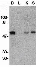
WB analysis of crude membrane fractions of human brain (B), liver (L), kidney (K), and spleen (S) using GTX11115 GDNF Receptor alpha 1 antibody. Working concentration : 1 μg/ml


WB analysis of crude membrane fractions of human brain (B), liver (L), kidney (K), and spleen (S) using GTX11115 GDNF Receptor alpha 1 antibody. Working concentration : 1 μg/ml
GDNF Receptor alpha 1 antibody
GTX11115
ApplicationsWestern Blot, ELISA, ImmunoHistoChemistry, ImmunoHistoChemistry Paraffin
Product group Antibodies
ReactivityHuman, Mouse, Rat
TargetGFRA1
Overview
- SupplierGeneTex
- Product NameGDNF Receptor alpha 1 antibody
- Delivery Days Customer9
- Application Supplier NoteWB: 1 microg/mL. IHC-P: 1 microg/mL. *Optimal dilutions/concentrations should be determined by the researcher.Not tested in other applications.
- ApplicationsWestern Blot, ELISA, ImmunoHistoChemistry, ImmunoHistoChemistry Paraffin
- CertificationResearch Use Only
- ClonalityPolyclonal
- Concentration1 mg/ml
- ConjugateUnconjugated
- Gene ID2674
- Target nameGFRA1
- Target descriptionGDNF family receptor alpha 1
- Target synonymsGDNFR, GDNFR-alpha-1, GDNFRA, GFR-ALPHA-1, GFRalpha-1, RET1L, RETL1, RHDA4, TRNR1, GDNF family receptor alpha-1, GDNF receptor alpha 1d, GDNF receptor alpha 1e, GPI-linked anchor protein, Glial cell line-derived neurotrophic factor receptor alpha, PI-linked cell-surface accessory protein, RET ligand 1, TGF-beta-related neurotrophic factor receptor 1
- HostRabbit
- IsotypeIgG
- Protein IDP56159
- Protein NameGDNF family receptor alpha-1
- Scientific DescriptionGlial cell line-derived neurotrophic factor (GDNF) and neurturin (NTN) are two structurally related, potent neurotrophic factors that play key roles in the control of neuron survival and differentiation. The protein encoded by this gene is a member of the GDNF receptor family. It is a glycosylphosphatidylinositol(GPI)-linked cell surface receptor for both GDNF and NTN, and mediates activation of the RET tyrosine kinase receptor. This gene is a candidate gene for Hirschsprung disease. Multiple alternatively spliced transcript variants have been described for this gene. [provided by RefSeq, Feb 2009]
- ReactivityHuman, Mouse, Rat
- Storage Instruction-20°C or -80°C,2°C to 8°C
- UNSPSC41116161
Datasheet
Related products
Product group Antibodies

GFR alpha 1 AntibodyABX013085
ApplicationsELISA, ImmunoHistoChemistry
- SizePrice
Product group Antibodies
Anti-GFRA1 Antibody144-65897
ApplicationsImmunoFluorescence, Western Blot
ReactivityHuman, Mouse, Rat
TargetGFRA1
- SizePrice
Product group Antibodies
Anti-GFRA1 AntibodyA35275
ApplicationsImmunoFluorescence, Western Blot, ImmunoHistoChemistry
ReactivityHuman, Mouse, Rat
- SizePrice
Product group Antibodies

GFRA1 / GFR Alpha AntibodyLS-C832593
ApplicationsWestern Blot, ELISA, ImmunoHistoChemistry
ReactivityHuman, Mouse, Rat
TargetGFRA1
- SizePrice
Product group Antibodies
References
GDNFRA Polyclonal AntibodyBS-0201R
ApplicationsFlow Cytometry, ImmunoFluorescence, Western Blot, ELISA, ImmunoCytoChemistry, ImmunoHistoChemistry, ImmunoHistoChemistry Frozen, ImmunoHistoChemistry Paraffin
ReactivityBovine, Canine, Equine, Human, Mouse, Porcine, Rat
TargetGFRA1
- SizePrice
Product group Antibodies
GFRA1 AntibodyCSB-PA008804
ApplicationsELISA, ImmunoHistoChemistry
ReactivityHuman, Mouse, Rat
TargetGFRA1
- SizePrice
Product group Antibodies
Gfra1 Polyclonal AntibodyCAC07821
ApplicationsImmunoFluorescence, Western Blot, ELISA, ImmunoHistoChemistry
ReactivityMouse
TargetGFRA1
- SizePrice

Product group Antibodies
Anti-GFRA1 AntibodyHPA043829
ApplicationsImmunoCytoChemistry, ImmunoHistoChemistry
ReactivityHuman
TargetGFRA1
- SizePrice
Product group Antibodies
GDNF Receptor alpha 1 antibodyGTX54146
ApplicationsImmunoFluorescence, Western Blot, ImmunoCytoChemistry, ImmunoHistoChemistry, ImmunoHistoChemistry Paraffin
ReactivityHuman, Mouse, Rat
TargetGFRA1
- SizePrice
Product group Antibodies
GDNF Receptor alpha 1 antibodyGTX41588
ApplicationsWestern Blot
ReactivityHuman, Mouse, Rat
TargetGFRA1
- SizePrice