

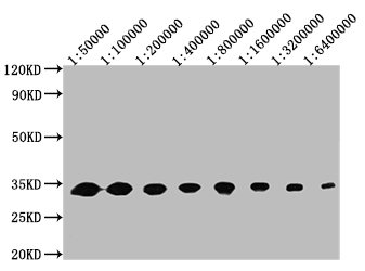
Western Blot Positive WB detected in: 50ng recombinant protein All lanes: GFP antibody at 1:50000, 1:100000, 1:200000, 1:400000, 1:800000, 1:1600000, 1:3200000, 1:6400000 Secondary Goat polyclonal to mouse IgG at 1/50000 dilution Predicted band size: 32 KDa Observed band size: 32 KDa Exposure time : 5min


.")
 or transfected with GFP (Right) stained with CSB-MA000051M1m at 1:200. The cells were fixed in 70% ethanol at 4°C overnight. Then 10% normal goat serum was Incubated to block non-specific protein-protein interactions followed by the antibody (1microg/1*106 cells) for 1 h at 4°C. The secondary antibody used was Alexa Fluor 647 AffiniPure Donkey Anti-Mouse IgG (H+L) at 1/250 dilution for 30min at 4°C.")
 or transfected with GFP (red line) stained with CSB-MA000051M1m at 1:200. The cells were fixed in 70% ethanol at 4°C overnight. Then 10% normal goat serum was Incubated to block non-specific protein-protein interactions followed by the antibody (1microg/1*106 cells) for 1 h at 4°C. The secondary antibody used was Alexa Fluor 647 AffiniPure Donkey Anti-Mouse IgG (H+L) at 1/250 dilution for 30min at 4°C.")
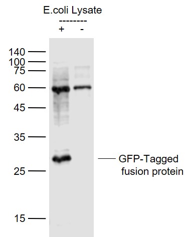
 + 293F whole cell lysate transfected with GFP (500microg) Lane 3: 293F whole cell lysate transfected with GFP (5microg) For western blotting, the blot was detected with CSB-MA000051M1m at 1:2000, and a HRP-conjugated Protein G antibody was used as the secondary antibody at 1:50000")
Western Blot Positive WB detected in: 50ng recombinant protein All lanes: GFP antibody at 1:50000, 1:100000, 1:200000, 1:400000, 1:800000, 1:1600000, 1:3200000, 1:6400000 Secondary Goat polyclonal to mouse IgG at 1/50000 dilution Predicted band size: 32 KDa Observed band size: 32 KDa Exposure time : 5min
GFP Monoclonal Antibody
CSB-MA000051M1M
ApplicationsFlow Cytometry, ImmunoFluorescence, ImmunoPrecipitation, Western Blot, ELISA
Product group Antibodies
ReactivityAll Species
Overview
- SupplierCusabio
- Product NameGFP Monoclonal Antibody
- Delivery Days Customer20
- ApplicationsFlow Cytometry, ImmunoFluorescence, ImmunoPrecipitation, Western Blot, ELISA
- CertificationResearch Use Only
- ClonalityMonoclonal
- Clone ID6C11C11
- ConjugateUnconjugated
- HostMouse
- IsotypeIgG2b
- Scientific DescriptionThe green fluorescent protein (GFP) is a protein that exhibit bright green fluorescence when exposed to blue light. The protein is in the shape of a cylinder, comprising 11 strands of beta-sheet with an alpha-helix inside and short helical segments on the ends of the cylinder. Inward-facing sidechains of the barrel induce specific cyclization reactions in the tripeptide Ser65-Tyr66-Gly67 that lead to chromophore formation. Its amazing ability to generate a highly visible, efficiently emitting internal fluorophore is both intrinsically fascinating and tremendously valuable. The green-fluorescent protein (GFP) of the jellyfish Aequorea victoria has always been used as a universal reporter in a broad range of heterologous living cells and organisms. GFP has become well established as a marker of gene expression and protein targeting in intact cells and organisms.
- ReactivityAll Species
- Storage Instruction-20°C or -80°C
- UNSPSC41116161
Related products
Product group Antibodies
GFP Monoclonal AntibodyCAC13721
ApplicationsFlow Cytometry, ImmunoFluorescence, ImmunoPrecipitation, Western Blot, ELISA
ReactivityOther Species
- SizePrice
Product group Antibodies
References
GFP Polyclonal AntibodyBS-0639R
ApplicationsImmunoFluorescence, Western Blot
ReactivityOther Species
TargetLOC107331116
- SizePrice
Product group Antibodies
GFP Monoclonal AntibodyMACO0649
ApplicationsImmunoPrecipitation, Western Blot, ELISA
- SizePrice
Product group Antibodies
GFP-Tag Monoclonal AntibodyCSB-MA000164
ApplicationsWestern Blot, ELISA
ReactivityAll Species
- SizePrice