

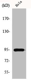
Western Blot analysis of HepG2 cells using GIT1 Polyclonal Antibody

Western Blot analysis of HepG2 cells using GIT1 Polyclonal Antibody
GIT1 Antibody
CSB-PA002688
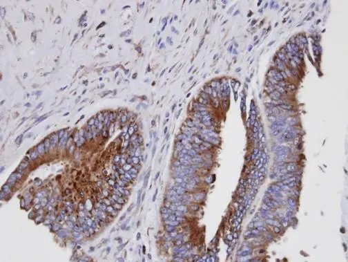
ApplicationsWestern Blot, ELISA, ImmunoHistoChemistry
Product group Antibodies
ReactivityHuman, Mouse, Rat
TargetGIT1
Overview
- SupplierCusabio
- Product NameGIT1 Antibody
- Delivery Days Customer20
- ApplicationsWestern Blot, ELISA, ImmunoHistoChemistry
- CertificationResearch Use Only
- ClonalityPolyclonal
- ConjugateUnconjugated
- Gene ID28964
- Target nameGIT1
- Target descriptionGIT ArfGAP 1
- Target synonymsp95-APP1, ARF GTPase-activating protein GIT1, ARF GAP GIT1, CAT-1, CAT1, G protein-coupled receptor kinase interacting ArfGAP 1, G protein-coupled receptor kinase-interactor 1, GRK-interacting protein 1, cool-associated and tyrosine-phosphorylated protein 1
- HostRabbit
- IsotypeIgG
- Protein IDQ9Y2X7
- Protein NameARF GTPase-activating protein GIT1
- ReactivityHuman, Mouse, Rat
- Storage Instruction-20°C or -80°C
- UNSPSC41116161
Related products

Product group Antibodies
Anti-GIT1 Antibody Picoband(r)A02140-3-CARRIER-FREE
ApplicationsFlow Cytometry, ImmunoFluorescence, Western Blot, ELISA, ImmunoCytoChemistry, ImmunoHistoChemistry
ReactivityHuman, Mouse, Rat
TargetGIT1
- SizePrice
Product group Antibodies
Anti-GIT1 Antibody144-61053
ApplicationsWestern Blot
ReactivityHuman, Mouse, Rat
TargetGIT1
- SizePrice

Product group Antibodies
Anti-GIT1 AntibodyA96195
ApplicationsWestern Blot, ELISA, ImmunoHistoChemistry
ReactivityHuman, Mouse, Rat
- SizePrice
Product group Antibodies

GIT1 AntibodyLS-C750405
ApplicationsWestern Blot
ReactivityHuman, Mouse, Rat
TargetGIT1
- SizePrice

Product group Antibodies
Anti-GIT1 AntibodyHPA004059
ApplicationsWestern Blot, ImmunoCytoChemistry, ImmunoHistoChemistry
ReactivityHuman
TargetGIT1
- SizePrice
Product group Antibodies
GIT1 antibody [N3C2], InternalGTX105824
ApplicationsImmunoFluorescence, Western Blot, ImmunoCytoChemistry, ImmunoHistoChemistry, ImmunoHistoChemistry Paraffin
ReactivityHuman, Mouse, Rat
TargetGIT1
- SizePrice
Product group Antibodies
GIT1 Recombinant AntibodyBSM-61866R
ApplicationsFlow Cytometry, Western Blot
ReactivityHuman, Mouse, Rat
TargetGIT1
- SizePrice
Product group Antibodies
GIT1 Polyclonal AntibodyCAC14837
ApplicationsImmunoFluorescence, Western Blot, ELISA, ImmunoHistoChemistry
ReactivityMouse
TargetGIT1
- SizePrice