

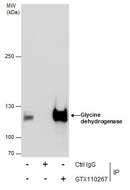
Immunoprecipitation of Glycine dehydrogenase protein from HepG2 whole cell extracts using 5 μg of Glycine dehydrogenase antibody [N3C2-2], Internal (GTX110267). Western blot analysis was performed using Glycine dehydrogenase antibody [N3C2-2], Internal (GTX110267). EasyBlot anti-Rabbit IgG (GTX221666-01) was used as a secondary reagent.
![Glycine dehydrogenase antibody [N3C2-2], Internal detects Glycine dehydrogenase protein at cytoplasm in mouse brain by immunohistochemical analysis. Sample: Paraffin-embedded mouse brain. Glycine dehydrogenase antibody [N3C2-2], Internal (GTX110267) diluted at 1:500.
Antigen Retrieval: Citrate buffer, pH 6.0, 15 min](https://www.genetex.com/upload/website/prouct_img/normal/GTX110267/GTX110267_42326_20160127_IHC-P_M_2_w_23060500_621.webp "Glycine dehydrogenase antibody [N3C2-2], Internal detects Glycine dehydrogenase protein at cytoplasm in mouse brain by immunohistochemical analysis. Sample: Paraffin-embedded mouse brain. Glycine dehydrogenase antibody [N3C2-2], Internal (GTX110267) diluted at 1:500.
Antigen Retrieval: Citrate buffer, pH 6.0, 15 min")
![Whole cell extract (30 μg) was separated by 5% SDS-PAGE, and the membranes were blotted with Glycine dehydrogenase antibody [N3C2-2], Internal (GTX110267) diluted at 1:1000 and competitor's antibody (CST#12794) diluted at 1:1000. The HRP-conjugated anti-rabbit IgG antibody (GTX213110-01) was used to detect the primary antibody.](https://www.genetex.com/upload/website/prouct_img/normal/GTX110267/GTX110267_42326_20171103_WB_competitor_watermark_w_23060500_761.webp "Whole cell extract (30 μg) was separated by 5% SDS-PAGE, and the membranes were blotted with Glycine dehydrogenase antibody [N3C2-2], Internal (GTX110267) diluted at 1:1000 and competitor's antibody (CST#12794) diluted at 1:1000. The HRP-conjugated anti-rabbit IgG antibody (GTX213110-01) was used to detect the primary antibody.")
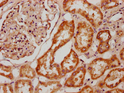
![Glycine dehydrogenase antibody [N3C2-2], Internal detects Glycine dehydrogenase protein at mitochondria in mouse liver by immunohistochemical analysis. Sample: Paraffin-embedded mouse liver. Glycine dehydrogenase antibody [N3C2-2], Internal (GTX110267) diluted at 1:500.
Antigen Retrieval: Citrate buffer, pH 6.0, 15 min](https://www.genetex.com/upload/website/prouct_img/normal/GTX110267/GTX110267_42326_20160127_IHC-P_M_1_w_23060500_822.webp "Glycine dehydrogenase antibody [N3C2-2], Internal detects Glycine dehydrogenase protein at mitochondria in mouse liver by immunohistochemical analysis. Sample: Paraffin-embedded mouse liver. Glycine dehydrogenase antibody [N3C2-2], Internal (GTX110267) diluted at 1:500.
Antigen Retrieval: Citrate buffer, pH 6.0, 15 min")
![Glycine dehydrogenase antibody [N3C2-2], Internal detects Glycine dehydrogenase protein at cytoplasm in rat brain by immunohistochemical analysis. Sample: Paraffin-embedded rat brain. Glycine dehydrogenase antibody [N3C2-2], Internal (GTX110267) diluted at 1:500.
Antigen Retrieval: Citrate buffer, pH 6.0, 15 min](https://www.genetex.com/upload/website/prouct_img/normal/GTX110267/GTX110267_42326_20160127_IHC-P_R_w_23060500_135.webp "Glycine dehydrogenase antibody [N3C2-2], Internal detects Glycine dehydrogenase protein at cytoplasm in rat brain by immunohistochemical analysis. Sample: Paraffin-embedded rat brain. Glycine dehydrogenase antibody [N3C2-2], Internal (GTX110267) diluted at 1:500.
Antigen Retrieval: Citrate buffer, pH 6.0, 15 min")
![Glycine dehydrogenase antibody [N3C2-2], Internal detects Glycine dehydrogenase protein at mitochondria by immunofluorescent analysis. Sample: Hep G2 cells were fixed in ice-cold MeOH for 5 min. Green: Glycine dehydrogenase protein stained by Glycine dehydrogenase antibody [N3C2-2], Internal (GTX110267) diluted at 1:400. Blue: Hoechst 33342 staining.](https://www.genetex.com/upload/website/prouct_img/normal/GTX110267/GTX110267_42326_20160411_IFA_w_23060500_770.webp "Glycine dehydrogenase antibody [N3C2-2], Internal detects Glycine dehydrogenase protein at mitochondria by immunofluorescent analysis. Sample: Hep G2 cells were fixed in ice-cold MeOH for 5 min. Green: Glycine dehydrogenase protein stained by Glycine dehydrogenase antibody [N3C2-2], Internal (GTX110267) diluted at 1:400. Blue: Hoechst 33342 staining.")
Immunoprecipitation of Glycine dehydrogenase protein from HepG2 whole cell extracts using 5 μg of Glycine dehydrogenase antibody [N3C2-2], Internal (GTX110267). Western blot analysis was performed using Glycine dehydrogenase antibody [N3C2-2], Internal (GTX110267). EasyBlot anti-Rabbit IgG (GTX221666-01) was used as a secondary reagent.
Glycine dehydrogenase antibody [N3C2-2], Internal
GTX110267
ApplicationsImmunoFluorescence, ImmunoPrecipitation, Western Blot, ImmunoCytoChemistry, ImmunoHistoChemistry, ImmunoHistoChemistry Paraffin
Product group Antibodies
ReactivityHuman, Mouse, Rat
TargetGLDC
Overview
- SupplierGeneTex
- Product NameGlycine dehydrogenase antibody [N3C2-2], Internal
- Delivery Days Customer9
- Application Supplier NoteWB: 1:500-1:3000. ICC/IF: 1:100-1:1000. IHC-P: 1:100-1:1000. IP: 1:100-1:500. *Optimal dilutions/concentrations should be determined by the researcher.Not tested in other applications.
- ApplicationsImmunoFluorescence, ImmunoPrecipitation, Western Blot, ImmunoCytoChemistry, ImmunoHistoChemistry, ImmunoHistoChemistry Paraffin
- CertificationResearch Use Only
- ClonalityPolyclonal
- Concentration0.46 mg/ml
- ConjugateUnconjugated
- Gene ID2731
- Target nameGLDC
- Target descriptionglycine decarboxylase
- Target synonymsGCE, GCE1, GCSP, HYGN1, glycine dehydrogenase (decarboxylating), mitochondrial, glycine cleavage system protein P, glycine decarboxylase P-protein, glycine dehydrogenase (aminomethyl-transferring), glycine dehydrogenase [decarboxylating], mitochondrial, nonketotic hyperglycinemia
- HostRabbit
- IsotypeIgG
- Protein IDP23378
- Protein NameGlycine dehydrogenase (decarboxylating), mitochondrial
- Scientific DescriptionThe enzyme system for cleavage of glycine (glycine cleavage system; GCS; EC 2.1.2.10), which is confined to the mitochondria, is composed of 4 protein components: P protein (a pyridoxal phosphate-dependent glycine decarboxylase), H protein (a lipoic acid-containing protein), T protein (a tetrahydrofolate-requiring enzyme), and L protein (a lipoamide dehydrogenase). Glycine encephalopathy (GCE; MIM 605899) may be due to a defect in any one of these enzymes; see MIM 238310, MIM 238330, and MIM 238331.[supplied by OMIM]
- ReactivityHuman, Mouse, Rat
- Storage Instruction-20°C or -80°C,2°C to 8°C
- UNSPSC41116161
Datasheet
Related products

Product group Antibodies
Anti-Glycine decarboxylase/GLDC Antibody Picoband(r)A04777-2-CARRIER-FREE
ApplicationsFlow Cytometry, Western Blot, ELISA, ImmunoHistoChemistry
ReactivityHuman, Mouse, Rat
TargetGLDC
- SizePrice
Product group Antibodies
Anti-GLDC Antibody144-09933
ApplicationsWestern Blot, ImmunoHistoChemistry
ReactivityHuman, Mouse
TargetGLDC
- SizePrice
Product group Antibodies
GLDC Polyclonal AntibodyBS-13370R
ApplicationsImmunoFluorescence, Western Blot, ELISA, ImmunoCytoChemistry, ImmunoHistoChemistry, ImmunoHistoChemistry Frozen, ImmunoHistoChemistry Paraffin
ReactivityCanine, Equine, Human, Mouse, Rabbit, Rat
TargetGLDC
- SizePrice
Product group Antibodies
GLDC AntibodyCSB-PA328400LA01HU
ApplicationsELISA, ImmunoHistoChemistry
ReactivityHuman
TargetGLDC
- SizePrice
Product group Antibodies
ApplicationsImmunoPrecipitation, Western Blot, ImmunoCytoChemistry, ImmunoHistoChemistry
ReactivityMouse, Rat
TargetGLDC
- SizePrice
Product group Antibodies

GLDC AntibodyLS-C411436
ApplicationsWestern Blot
ReactivityHuman, Mouse
TargetGLDC
- SizePrice

Product group Antibodies
Anti-GLDC AntibodyHPA002318
ApplicationsWestern Blot, ImmunoHistoChemistry
ReactivityHuman, Mouse, Rat
TargetGLDC
- SizePrice
Product group Antibodies
Anti-GLDC AntibodyCAB9933
ApplicationsImmunoFluorescence, ImmunoPrecipitation, Western Blot, ELISA, ImmunoCytoChemistry
ReactivityHuman
TargetGLDC
- SizePrice