

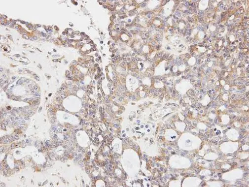
Immunohistochemical analysis of paraffin-embedded OVCA xenograft, using GLYCTK(GTX107807) antibody at 1:100 dilution.
Antigen Retrieval: Trilogy? (EDTA based, pH 8.0) buffer, 15min
![GLYCTK antibody [N3C3] detects GLYCTK protein at cytoplasm by confocal immunofluorescent analysis. Sample: HepG2 cells were fixed in 2% paraformaldehyde/culture medium at 37oC for 30 min. Green: GLYCTK protein stained by GLYCTK antibody [N3C3] (GTX107807) diluted at 1:500. Blue: Hoechst 33342 staining. [Images captured by Olympus FV10i Confocal Laser Scanning Microscope]](https://www.genetex.com/upload/website/prouct_img/normal/GTX107807/GTX107807_40317_IFA_w_23060120_623.webp "GLYCTK antibody [N3C3] detects GLYCTK protein at cytoplasm by confocal immunofluorescent analysis. Sample: HepG2 cells were fixed in 2% paraformaldehyde/culture medium at 37oC for 30 min. Green: GLYCTK protein stained by GLYCTK antibody [N3C3] (GTX107807) diluted at 1:500. Blue: Hoechst 33342 staining. [Images captured by Olympus FV10i Confocal Laser Scanning Microscope]")
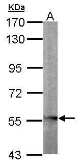
 A:A431(GTX27909) B:Hep G2(GTX27900) 7.5% SDS PAGE GTX107807 diluted at 1:500")
Immunohistochemical analysis of paraffin-embedded OVCA xenograft, using GLYCTK(GTX107807) antibody at 1:100 dilution.
Antigen Retrieval: Trilogy? (EDTA based, pH 8.0) buffer, 15min
GLYCTK antibody [N3C3]
GTX107807
ApplicationsImmunoFluorescence, Western Blot, ImmunoCytoChemistry, ImmunoHistoChemistry, ImmunoHistoChemistry Paraffin
Product group Antibodies
ReactivityHuman
TargetGLYCTK
Overview
- SupplierGeneTex
- Product NameGLYCTK antibody [N3C3]
- Delivery Days Customer9
- Application Supplier NoteWB: 1:500-1:3000. ICC/IF: 1:100-1:1000. IHC-P: 1:100-1:1000. *Optimal dilutions/concentrations should be determined by the researcher.Not tested in other applications.
- ApplicationsImmunoFluorescence, Western Blot, ImmunoCytoChemistry, ImmunoHistoChemistry, ImmunoHistoChemistry Paraffin
- CertificationResearch Use Only
- ClonalityPolyclonal
- Concentration0.54 mg/ml
- ConjugateUnconjugated
- Gene ID132158
- Target nameGLYCTK
- Target descriptionglycerate kinase
- Target synonymsHBEBP2, HBEBP4, HBeAgBP4A, glycerate kinase, HBeAg binding protein 4, HBeAg-binding protein 2
- HostRabbit
- IsotypeIgG
- Protein IDQ8IVS8
- Protein NameGlycerate kinase
- Scientific DescriptionThis locus encodes a member of the glycerate kinase type-2 family. The encoded enzyme catalyzes the phosphorylation of (R)-glycerate and may be involved in serine degradation and fructose metabolism. Decreased activity of the encoded enzyme may be associated with the disease D-glyceric aciduria. Alternatively spliced transcript variants have been described. [provided by RefSeq]
- ReactivityHuman
- Storage Instruction-20°C or -80°C,2°C to 8°C
- UNSPSC41116161
Datasheet
Related products
Product group Antibodies
GLYCTK AntibodyCSB-PA007152
ApplicationsImmunoFluorescence, Western Blot, ELISA, ImmunoHistoChemistry
ReactivityHuman, Mouse, Rat
TargetGLYCTK
- SizePrice

Product group Antibodies
ApplicationsImmunoFluorescence, Western Blot, ELISA, ImmunoCytoChemistry, ImmunoHistoChemistry
TargetGLYCTK
- SizePrice

Product group Antibodies
Anti-GLYCTK-25ulHPA006913
ApplicationsWestern Blot, ImmunoHistoChemistry
ReactivityHuman, Mouse
- SizePrice
Product group Antibodies

ApplicationsImmunoFluorescence, Western Blot, ImmunoCytoChemistry, ImmunoHistoChemistry, ImmunoHistoChemistry Paraffin
ReactivityBovine, Human, Monkey, Mouse, Rat
TargetGLYCTK
- SizePrice
Product group Antibodies
GLYCTK antibody [N2C3]GTX103789
ApplicationsWestern Blot
ReactivityHuman, Mouse
TargetGLYCTK
- SizePrice
Product group Antibodies
GLYCTK antibodyGTX33222
ApplicationsWestern Blot
ReactivityHuman, Mouse, Rat
TargetGLYCTK
- SizePrice
Product group Antibodies
Anti-GLYCTK Antibody144-07852
ApplicationsWestern Blot
ReactivityHuman, Mouse, Rat
TargetGLYCTK
- SizePrice
Product group Antibodies
GLYCTK Polyclonal AntibodyBS-13448R
ApplicationsImmunoFluorescence, Western Blot, ELISA, ImmunoCytoChemistry, ImmunoHistoChemistry, ImmunoHistoChemistry Frozen, ImmunoHistoChemistry Paraffin
ReactivityHuman, Mouse, Rat
TargetGLYCTK
- SizePrice