

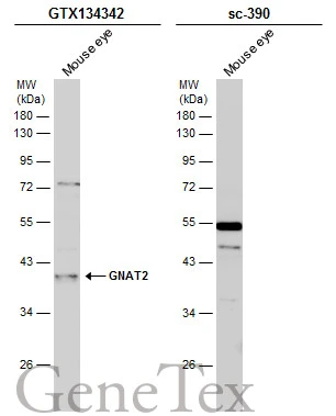
Mouse tissue extract (50 μg) was separated by 10% SDS-PAGE, and the membranes were blotted with GNAT2 antibody (GTX134342) diluted at 1:1000 and competitor's antibody (sc-390) diluted at 1:1000. The HRP-conjugated anti-rabbit IgG antibody (GTX213110-01) was used to detect the primary antibody. *The competitor is not affiliated with GeneTex and does not endorse this product.
![GNAT2 antibody detects GNAT2 protein at cell membrane and cytoplasm by immunohistochemical analysis. Sample: Paraffin-embedded mouse eye. Green: GNAT2 stained by GNAT2 antibody (GTX134342) diluted at 1:200. Red: beta Tubulin 3/ Tuj1, a cytoskeleton marker, stained by beta Tubulin 3/ Tuj1 antibody [GT1338] (GTX631831) diluted at 1:500. Blue: Fluoroshield with DAPI (GTX30920).
Antigen Retrieval: Citrate buffer, pH 6.0, 15 min](https://www.genetex.com/upload/website/prouct_img/normal/GTX134342/GTX134342_43173_20180504_IHC-P-FL_M_w_23060620_124.webp "GNAT2 antibody detects GNAT2 protein at cell membrane and cytoplasm by immunohistochemical analysis. Sample: Paraffin-embedded mouse eye. Green: GNAT2 stained by GNAT2 antibody (GTX134342) diluted at 1:200. Red: beta Tubulin 3/ Tuj1, a cytoskeleton marker, stained by beta Tubulin 3/ Tuj1 antibody [GT1338] (GTX631831) diluted at 1:500. Blue: Fluoroshield with DAPI (GTX30920).
Antigen Retrieval: Citrate buffer, pH 6.0, 15 min")
![GNAT2 antibody detects GNAT2 protein by immunofluorescent analysis. Sample: DIV9 rat E18 primary cortical neuron cells were fixed in 4% paraformaldehyde at RT for 15 min. Green: GNAT2 stained by GNAT2 antibody (GTX134342) diluted at 1:500. Red: Tau, stained by Tau antibody [GT287] (GTX634809) diluted at 1:500. Blue: Fluoroshield with DAPI (GTX30920).](https://www.genetex.com/upload/website/prouct_img/normal/GTX134342/GTX134342_43173_20190306_ICC_IF_R_w_23060620_372.webp "GNAT2 antibody detects GNAT2 protein by immunofluorescent analysis. Sample: DIV9 rat E18 primary cortical neuron cells were fixed in 4% paraformaldehyde at RT for 15 min. Green: GNAT2 stained by GNAT2 antibody (GTX134342) diluted at 1:500. Red: Tau, stained by Tau antibody [GT287] (GTX634809) diluted at 1:500. Blue: Fluoroshield with DAPI (GTX30920).")
Mouse tissue extract (50 μg) was separated by 10% SDS-PAGE, and the membranes were blotted with GNAT2 antibody (GTX134342) diluted at 1:1000 and competitor's antibody (sc-390) diluted at 1:1000. The HRP-conjugated anti-rabbit IgG antibody (GTX213110-01) was used to detect the primary antibody. *The competitor is not affiliated with GeneTex and does not endorse this product.
GNAT2 antibody
GTX134342
ApplicationsImmunoFluorescence, Western Blot, ImmunoCytoChemistry, ImmunoHistoChemistry, ImmunoHistoChemistry Paraffin
Product group Antibodies
ReactivityHuman, Mouse, Rat
TargetGNAT2
Overview
- SupplierGeneTex
- Product NameGNAT2 antibody
- Delivery Days Customer9
- Application Supplier NoteWB: 1:500-1:3000. ICC/IF: 1:100-1:1000. IHC-P: 1:100-1:1000. *Optimal dilutions/concentrations should be determined by the researcher.Not tested in other applications.
- ApplicationsImmunoFluorescence, Western Blot, ImmunoCytoChemistry, ImmunoHistoChemistry, ImmunoHistoChemistry Paraffin
- CertificationResearch Use Only
- ClonalityPolyclonal
- Concentration0.92 mg/ml
- ConjugateUnconjugated
- Gene ID2780
- Target nameGNAT2
- Target descriptionG protein subunit alpha transducin 2
- Target synonymsACHM4, GNATC, HG1D, guanine nucleotide-binding protein G(t) subunit alpha-2, cone-type transducin alpha subunit, guanine nucleotide binding protein (G protein), alpha transducing activity polypeptide 2, heterotrimeric guanine nucleotide-binding protein 1D, transducin alpha-2 chain, transducin, cone-specific, alpha polypeptide
- HostRabbit
- IsotypeIgG
- Protein IDP19087
- Protein NameGuanine nucleotide-binding protein G(t) subunit alpha-2
- Scientific DescriptionTransducin is a 3-subunit guanine nucleotide-binding protein (G protein) which stimulates the coupling of rhodopsin and cGMP-phoshodiesterase during visual impulses. The transducin alpha subunits in rods and cones are encoded by separate genes. This gene encodes the alpha subunit in cones. [provided by RefSeq, Jul 2008]
- ReactivityHuman, Mouse, Rat
- Storage Instruction-20°C or -80°C,2°C to 8°C
- UNSPSC41116161
Datasheet
Related products
Product group Antibodies
GNAT2 AntibodyCSB-PA008888
ApplicationsWestern Blot, ELISA, ImmunoHistoChemistry
ReactivityHuman, Mouse
TargetGNAT2
- SizePrice
Product group Antibodies
ApplicationsWestern Blot, ImmunoHistoChemistry
ReactivityHuman, Mouse
TargetGNAT2
- SizePrice
Product group Antibodies
Anti-GNAT2 AntibodyA99088
ApplicationsWestern Blot, ELISA
ReactivityHuman, Mouse
- SizePrice
Product group Antibodies

GNAT2 AntibodyLS-C497061
ApplicationsWestern Blot
ReactivityHuman
TargetGNAT2
- SizePrice
![GNAT2 antibody [N3C3] detects GNAT2 protein expression by immunohistochemical analysis. Sample: Frozen sectioned adult mouse retina. Green: GNAT2 protein stained by GNAT2 antibody [N3C3] (GTX114440) diluted at 1:250. Red: beta Tubulin 3/ TUJ1, stained by beta Tubulin 3/ TUJ1 antibody [GT11710] (GTX631836) diluted at 1:250. Blue: Fluoroshield with DAPI (GTX30920).](https://www.genetex.com/upload/website/prouct_img/normal/GTX114440/GTX114440_41808_20170214_IHC-Fr_w_23060501_362.webp)
Product group Antibodies
GNAT2 antibody [N3C3]GTX114440
ApplicationsImmunoHistoChemistry, ImmunoHistoChemistry Frozen
ReactivityHuman, Mouse
TargetGNAT2
- SizePrice
Product group Antibodies
Anti-GNAT2 (Center) Antibody102-20729
ApplicationsWestern Blot, ImmunoHistoChemistry, ImmunoHistoChemistry Paraffin
TargetGNAT2
- SizePrice