

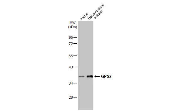
HeLa whole cell and nuclear extracts (30 μg) were separated by 10% SDS-PAGE, and the membrane was blotted with GPS2 antibody [HL2276] (GTX638329) diluted at 1:1000. The HRP-conjugated anti-rabbit IgG antibody (GTX213110-01) was used to detect the primary antibody, and the signal was developed with Trident ECL plus-Enhanced.
![Whole zebrafish extract (30 μg) was separated by 10% SDS-PAGE, and the membrane was blotted with GPS2 antibody [HL2276] (GTX638329) diluted at 1:1000. The HRP-conjugated anti-rabbit IgG antibody (GTX213110-01) was used to detect the primary antibody, and the signal was developed with Trident ECL plus-Enhanced.](https://www.genetex.com/upload/website/prouct_img/normal/GTX638329/GTX638329_45075_20230616_WB_Z_23062718_932.webp "Whole zebrafish extract (30 μg) was separated by 10% SDS-PAGE, and the membrane was blotted with GPS2 antibody [HL2276] (GTX638329) diluted at 1:1000. The HRP-conjugated anti-rabbit IgG antibody (GTX213110-01) was used to detect the primary antibody, and the signal was developed with Trident ECL plus-Enhanced.")
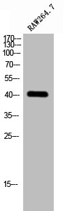
![Various tissue extracts (30 μg) were separated by 10% SDS-PAGE, and the membrane was blotted with GPS2 antibody [HL2276] (GTX638329) diluted at 1:1000. The HRP-conjugated anti-rabbit IgG antibody (GTX213110-01) was used to detect the primary antibody.](https://www.genetex.com/upload/website/prouct_img/normal/GTX638329/GTX638329_45075_20230825_WB_M_tissue_23083020_403.webp "Various tissue extracts (30 μg) were separated by 10% SDS-PAGE, and the membrane was blotted with GPS2 antibody [HL2276] (GTX638329) diluted at 1:1000. The HRP-conjugated anti-rabbit IgG antibody (GTX213110-01) was used to detect the primary antibody.")
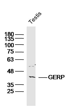
![Rat tissue extract (50 μg) was separated by 10% SDS-PAGE, and the membrane was blotted with GPS2 antibody [HL2276] (GTX638329) diluted at 1:10000. The HRP-conjugated anti-rabbit IgG antibody (GTX213110-01) was used to detect the primary antibody.](https://www.genetex.com/upload/website/prouct_img/normal/GTX638329/GTX638329_45075_20230901_WB_R_muscle_23090619_421.webp "Rat tissue extract (50 μg) was separated by 10% SDS-PAGE, and the membrane was blotted with GPS2 antibody [HL2276] (GTX638329) diluted at 1:10000. The HRP-conjugated anti-rabbit IgG antibody (GTX213110-01) was used to detect the primary antibody.")
![GPS2 antibody [HL2276] detects GPS2 protein at mitochondria by immunohistochemical analysis. Sample: Paraffin-embedded mouse salivary gland. GPS2 stained by GPS2 antibody [HL2276] (GTX638329) diluted at 1:1000. Antigen Retrieval: Citrate buffer, pH 6.0, 15 min](https://www.genetex.com/upload/website/prouct_img/normal/GTX638329/GTX638329_45075_20230829_IHC-P_M_23091319_715.webp "GPS2 antibody [HL2276] detects GPS2 protein at mitochondria by immunohistochemical analysis. Sample: Paraffin-embedded mouse salivary gland. GPS2 stained by GPS2 antibody [HL2276] (GTX638329) diluted at 1:1000. Antigen Retrieval: Citrate buffer, pH 6.0, 15 min")
![GPS2 antibody [HL2276] detects GPS2 protein by immunohistochemical analysis. Sample: Paraffin-embedded rat testis. GPS2 stained by GPS2 antibody [HL2276] (GTX638329) diluted at 1:1000. Antigen Retrieval: Citrate buffer, pH 6.0, 15 min](https://www.genetex.com/upload/website/prouct_img/normal/GTX638329/GTX638329_45075_20230829_IHC-P_R_23091319_320.webp "GPS2 antibody [HL2276] detects GPS2 protein by immunohistochemical analysis. Sample: Paraffin-embedded rat testis. GPS2 stained by GPS2 antibody [HL2276] (GTX638329) diluted at 1:1000. Antigen Retrieval: Citrate buffer, pH 6.0, 15 min")
![GPS2 antibody [HL2276] detects GPS2 protein at cytoplasm by immunofluorescent analysis. Sample: 293T cells were fixed in ice-cold MeOH for 5 min. Green: GPS2 stained by GPS2 antibody [HL2276] (GTX638329) diluted at 1:500. Blue: Fluoroshield with DAPI (GTX30920).](https://www.genetex.com/upload/website/prouct_img/normal/GTX638329/GTX638329_45075_20231006_ICC_IF_23102401_168.webp "GPS2 antibody [HL2276] detects GPS2 protein at cytoplasm by immunofluorescent analysis. Sample: 293T cells were fixed in ice-cold MeOH for 5 min. Green: GPS2 stained by GPS2 antibody [HL2276] (GTX638329) diluted at 1:500. Blue: Fluoroshield with DAPI (GTX30920).")
HeLa whole cell and nuclear extracts (30 μg) were separated by 10% SDS-PAGE, and the membrane was blotted with GPS2 antibody [HL2276] (GTX638329) diluted at 1:1000. The HRP-conjugated anti-rabbit IgG antibody (GTX213110-01) was used to detect the primary antibody, and the signal was developed with Trident ECL plus-Enhanced.
GPS2 antibody [HL2276]
GTX638329
ApplicationsImmunoFluorescence, Western Blot, ImmunoCytoChemistry, ImmunoHistoChemistry, ImmunoHistoChemistry Paraffin
Product group Antibodies
ReactivityHuman, Mouse, Rat, Zebra Fish
TargetGPS2
Overview
- SupplierGeneTex
- Product NameGPS2 antibody [HL2276]
- Delivery Days Customer9
- Application Supplier NoteWB: 1:500-1:3000. *Optimal dilutions/concentrations should be determined by the researcher.Not tested in other applications.
- ApplicationsImmunoFluorescence, Western Blot, ImmunoCytoChemistry, ImmunoHistoChemistry, ImmunoHistoChemistry Paraffin
- CertificationResearch Use Only
- ClonalityMonoclonal
- Clone IDHL2276
- Concentration2 mg/ml
- ConjugateUnconjugated
- Gene ID2874
- Target nameGPS2
- Target descriptionG protein pathway suppressor 2
- Target synonymsAMF-1, G protein pathway suppressor 2, GPS-2
- HostRabbit
- IsotypeIgG
- Protein IDQ13227
- Protein NameG protein pathway suppressor 2
- Scientific DescriptionThis gene encodes a protein involved in G protein-mitogen-activated protein kinase (MAPK) signaling cascades. When overexpressed in mammalian cells, this gene could potently suppress a RAS- and MAPK-mediated signal and interfere with JNK activity, suggesting that the function of this gene may be signal repression. The encoded protein is an integral subunit of the NCOR1-HDAC3 (nuclear receptor corepressor 1-histone deacetylase 3) complex, and it was shown that the complex inhibits JNK activation through this subunit and thus could potentially provide an alternative mechanism for hormone-mediated antagonism of AP1 (activator protein 1) function. [provided by RefSeq, Jul 2008]
- ReactivityHuman, Mouse, Rat, Zebra Fish
- Storage Instruction-20°C or -80°C,2°C to 8°C
- UNSPSC12352203
Datasheet
Related products

Product group Antibodies
Anti-GPS2 AntibodyA06569
ApplicationsWestern Blot, ELISA
ReactivityHuman, Mouse, Rat
TargetGPS2
- SizePrice
Product group Antibodies
GPS2 Polyclonal AntibodyBS-15395R
ApplicationsFlow Cytometry, ImmunoFluorescence, Western Blot, ELISA, ImmunoCytoChemistry, ImmunoHistoChemistry, ImmunoHistoChemistry Frozen, ImmunoHistoChemistry Paraffin
ReactivityBovine, Canine, Human, Mouse, Porcine, Rat, Sheep
- SizePrice
Product group Antibodies
GPS2 AntibodyCSB-PA008949
ApplicationsWestern Blot, ELISA
ReactivityHuman, Mouse, Rat
TargetGPS2
- SizePrice

Product group Antibodies
Anti-GPS2 AntibodyHPA067540
ApplicationsImmunoCytoChemistry, ImmunoHistoChemistry
ReactivityHuman
TargetGPS2
- SizePrice
Product group Antibodies

GPS2 AntibodyLS-C667801
ApplicationsWestern Blot
ReactivityHuman
TargetGPS2
- SizePrice
Product group Antibodies
References
GPS2 antibodyGTX117560
ApplicationsImmunoFluorescence, Western Blot, ImmunoCytoChemistry, ImmunoHistoChemistry, ImmunoHistoChemistry Paraffin
ReactivityHuman
TargetGPS2
- SizePrice

Product group Antibodies
Anti-GPS2 AntibodyA98118
ApplicationsWestern Blot, ELISA
ReactivityHuman, Mouse, Rat
- SizePrice