

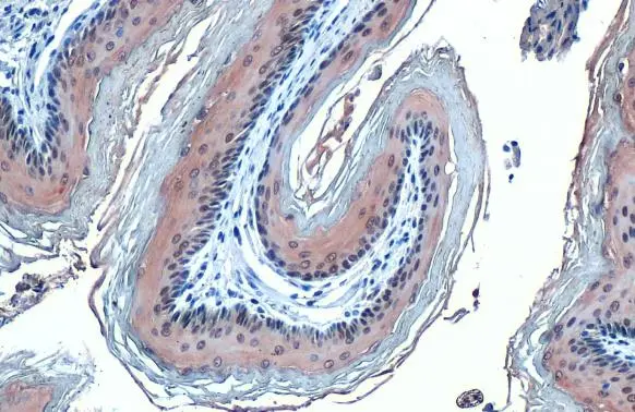
GPX2 antibody [C1C3] detects GPX2 protein at cytoplasm by immunohistochemical analysis. Sample: Paraffin-embedded mouse esophagus. GPX2 stained by GPX2 antibody [C1C3] (GTX100292) diluted at 1:500. Antigen Retrieval: Citrate buffer, pH 6.0, 15 min
![GPX2 antibody [C1C3] detects GPX2 protein at cytoplasm by immunohistochemical analysis. Sample: Paraffin-embedded rat liver. GPX2 stained by GPX2 antibody [C1C3] (GTX100292) diluted at 1:500. Antigen Retrieval: Citrate buffer, pH 6.0, 15 min](https://www.genetex.com/upload/website/prouct_img/normal/GTX100292/GTX100292_44741_20220805_IHC-P_R_22081423_787.webp "GPX2 antibody [C1C3] detects GPX2 protein at cytoplasm by immunohistochemical analysis. Sample: Paraffin-embedded rat liver. GPX2 stained by GPX2 antibody [C1C3] (GTX100292) diluted at 1:500. Antigen Retrieval: Citrate buffer, pH 6.0, 15 min")
![GPX2 antibody [C1C3] detects GPX2 protein at cytoplasm by immunohistochemical analysis. Sample: Paraffin-embedded mouse intestine. GPX2 stained by GPX2 antibody [C1C3] (GTX100292) diluted at 1:500. Antigen Retrieval: Citrate buffer, pH 6.0, 15 min](https://www.genetex.com/upload/website/prouct_img/normal/GTX100292/GTX100292_44741_20220805_IHC-P_M_2_22081423_978.webp "GPX2 antibody [C1C3] detects GPX2 protein at cytoplasm by immunohistochemical analysis. Sample: Paraffin-embedded mouse intestine. GPX2 stained by GPX2 antibody [C1C3] (GTX100292) diluted at 1:500. Antigen Retrieval: Citrate buffer, pH 6.0, 15 min")
![GPX2 antibody [C1C3] detects GPX2 protein at cytoplasm by immunohistochemical analysis. Sample: Paraffin-embedded rat duodenum. GPX2 stained by GPX2 antibody [C1C3] (GTX100292) diluted at 1:500. Antigen Retrieval: Citrate buffer, pH 6.0, 15 min](https://www.genetex.com/upload/website/prouct_img/normal/GTX100292/GTX100292_44776_20220909_IHC-P_R_22101319_756.webp "GPX2 antibody [C1C3] detects GPX2 protein at cytoplasm by immunohistochemical analysis. Sample: Paraffin-embedded rat duodenum. GPX2 stained by GPX2 antibody [C1C3] (GTX100292) diluted at 1:500. Antigen Retrieval: Citrate buffer, pH 6.0, 15 min")
![GPX2 antibody [C1C3] detects GPX2 protein at cytoplasm and nucleus by immunohistochemical analysis. Sample: Paraffin-embedded mouse stomach. GPX2 stained by GPX2 antibody [C1C3] (GTX100292) diluted at 1:500. Antigen Retrieval: Citrate buffer, pH 6.0, 15 min](https://www.genetex.com/upload/website/prouct_img/normal/GTX100292/GTX100292_44776_20220909_IHC-P_M_22101319_412.webp "GPX2 antibody [C1C3] detects GPX2 protein at cytoplasm and nucleus by immunohistochemical analysis. Sample: Paraffin-embedded mouse stomach. GPX2 stained by GPX2 antibody [C1C3] (GTX100292) diluted at 1:500. Antigen Retrieval: Citrate buffer, pH 6.0, 15 min")
![Immunoprecipitation of GPX2 protein from HepG2 whole cell extracts using 5 μg of GPX2 antibody [C1C3] (GTX100292). Western blot analysis was performed using GPX2 antibody [C1C3] (GTX100292). EasyBlot anti-Rabbit IgG (GTX221666-01) was used as a secondary reagent.](https://www.genetex.com/upload/website/prouct_img/normal/GTX100292/GTX100292_39428_20150430_IP_w_23060100_386.webp "Immunoprecipitation of GPX2 protein from HepG2 whole cell extracts using 5 μg of GPX2 antibody [C1C3] (GTX100292). Western blot analysis was performed using GPX2 antibody [C1C3] (GTX100292). EasyBlot anti-Rabbit IgG (GTX221666-01) was used as a secondary reagent.")
 antibody at 1:500 dilution.
Antigen Retrieval: Trilogy? (EDTA based, pH 8.0) buffer, 15min")
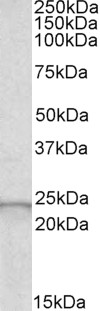
![Non-transfected (–) and transfected (+) HepG2 whole cell extracts (30 μg) were separated by 12% SDS-PAGE, and the membrane was blotted with GPX2 antibody [C1C3] (GTX100292) diluted at 1:10000. The HRP-conjugated anti-rabbit IgG antibody (GTX213110-01) was used to detect the primary antibody.](https://www.genetex.com/upload/website/prouct_img/normal/GTX100292/GTX100292_43985_20211210_WB_shRNA_watermark_w_23060100_168.webp "Non-transfected (–) and transfected (+) HepG2 whole cell extracts (30 μg) were separated by 12% SDS-PAGE, and the membrane was blotted with GPX2 antibody [C1C3] (GTX100292) diluted at 1:10000. The HRP-conjugated anti-rabbit IgG antibody (GTX213110-01) was used to detect the primary antibody.")
 was separated by 12 % SDS-PAGE, and blotted with GPX2 antibody (GTX100292) diluted by 1:500")
 antibody at 1:500 dilution.
Antigen Retrieval: Trilogy? (EDTA based, pH 8.0) buffer, 15min")
GPX2 antibody [C1C3] detects GPX2 protein at cytoplasm by immunohistochemical analysis. Sample: Paraffin-embedded mouse esophagus. GPX2 stained by GPX2 antibody [C1C3] (GTX100292) diluted at 1:500. Antigen Retrieval: Citrate buffer, pH 6.0, 15 min
GPX2 antibody [C1C3]
GTX100292
ApplicationsImmunoFluorescence, ImmunoPrecipitation, Western Blot, ImmunoCytoChemistry, ImmunoHistoChemistry, ImmunoHistoChemistry Paraffin
Product group Antibodies
ReactivityHuman, Mouse, Rat
TargetGPX2
Overview
- SupplierGeneTex
- Product NameGPX2 antibody [C1C3]
- Delivery Days Customer9
- Application Supplier NoteWB: 1:500-1:10000. IHC-P: 1:100-1:1000. IP: 1:100-1:500. *Optimal dilutions/concentrations should be determined by the researcher.Not tested in other applications.
- ApplicationsImmunoFluorescence, ImmunoPrecipitation, Western Blot, ImmunoCytoChemistry, ImmunoHistoChemistry, ImmunoHistoChemistry Paraffin
- CertificationResearch Use Only
- ClonalityPolyclonal
- Concentration1.37 mg/ml
- ConjugateUnconjugated
- Gene ID2877
- Target nameGPX2
- Target descriptionglutathione peroxidase 2
- Target synonymsGI-GPx, GPRP, GPRP-2, GPx-2, GPx-GI, GSHPX-GI, GSHPx-2, glutathione peroxidase 2, gastrointestinal glutathione peroxidase, glutathione peroxidase 2 (gastrointestinal), glutathione peroxidase-related protein 2, phospholipid hydroperoxide glutathione peroxidase GPX2, selenoprotein GPX2
- HostRabbit
- IsotypeIgG
- Protein IDP18283
- Protein NameGlutathione peroxidase 2
- Scientific DescriptionThis gene is a member of the glutathione peroxidase family and encodes a selenium-dependent glutathione peroxidase that is one of two isoenzymes responsible for the majority of the glutathione-dependent hydrogen peroxide-reducing activity in the epithelium of the gastrointestinal tract. Studies in knockout mice indicate that mRNA expression levels respond to luminal microflora, suggesting a role of the ileal glutathione peroxidases in preventing inflammation in the GI tract. [provided by RefSeq]
- ReactivityHuman, Mouse, Rat
- Storage Instruction-20°C or -80°C,2°C to 8°C
- UNSPSC41116161
Datasheet
Related products
Product group Antibodies
ApplicationsWestern Blot, ELISA
ReactivityHuman
- SizePrice
Product group Antibodies
Anti-GPX2 Antibody144-64016
ApplicationsWestern Blot, ImmunoHistoChemistry
ReactivityHuman, Mouse, Rat
TargetGPX2
- SizePrice
Product group Antibodies

GPX2 Antibody (Biotin)LS-C682198
ApplicationsELISA
ReactivityHuman
TargetGPX2
- SizePrice
Product group Antibodies
GPX2 Polyclonal AntibodyBS-13396R
ApplicationsImmunoFluorescence, Western Blot, ELISA, ImmunoCytoChemistry, ImmunoHistoChemistry, ImmunoHistoChemistry Frozen, ImmunoHistoChemistry Paraffin
ReactivityBovine, Canine, Equine, Human, Mouse, Rabbit, Rat, Sheep
TargetGPX2
- SizePrice
Product group Antibodies
GPX2 AntibodyCSB-PA009867LA01HU
ApplicationsImmunoFluorescence, ELISA
ReactivityHuman
TargetGPX2
- SizePrice
Product group Antibodies

ApplicationsWestern Blot, ELISA
ReactivityBovine, Canine, Human, Mouse, Porcine, Rat
TargetGPX2
- SizePrice
Product group Antibodies
GPX2 Polyclonal AntibodyCAC13053
ApplicationsImmunoFluorescence, ELISA
TargetGPX2
- SizePrice

Product group Antibodies
ApplicationsWestern Blot, ImmunoHistoChemistry
ReactivityHuman
TargetGPX2
- SizePrice

Product group Antibodies
Anti-GPX2 AntibodyHPA003545
ApplicationsImmunoHistoChemistry
ReactivityHuman
TargetGPX2
- SizePrice
![IHC-P analysis of human mucosa from descending colon tissue using GTX639924 GPX2 antibody [HMV301] HistoMAX?. Strong predominantly cytoplasmic GPX2 staining of epithelial cells - predominantly in the crypts. A luminal membrane staining can also be seen.](https://www.genetex.com/upload/website/prouct_img/normal/GTX639924/GTX639924_20240403_IHC-P_2_24040301_403.webp)
Product group Antibodies
GPX2 antibody [HMV301] HistoMAX(tm)GTX639924
ApplicationsImmunoHistoChemistry, ImmunoHistoChemistry Paraffin
ReactivityHuman
TargetGPX2
- SizePrice