

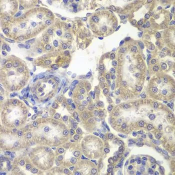
IHC-P analysis of rat kidney tissue using GTX54162 H6PD antibody. Dilution : 1:200


IHC-P analysis of rat kidney tissue using GTX54162 H6PD antibody. Dilution : 1:200
H6PD antibody
GTX54162
ApplicationsImmunoFluorescence, Western Blot, ImmunoCytoChemistry, ImmunoHistoChemistry, ImmunoHistoChemistry Paraffin
Product group Antibodies
ReactivityHuman, Mouse, Rat
TargetH6PD
Overview
- SupplierGeneTex
- Product NameH6PD antibody
- Delivery Days Customer7
- Application Supplier NoteWB: 1:500 - 1:2000. ICC/IF: 1:10 - 1:100. IHC-P: 1:50 - 1:200. *Optimal dilutions/concentrations should be determined by the researcher.Not tested in other applications.
- ApplicationsImmunoFluorescence, Western Blot, ImmunoCytoChemistry, ImmunoHistoChemistry, ImmunoHistoChemistry Paraffin
- CertificationResearch Use Only
- ClonalityPolyclonal
- Concentration1 mg/ml
- ConjugateUnconjugated
- Gene ID9563
- Target nameH6PD
- Target descriptionhexose-6-phosphate dehydrogenase/glucose 1-dehydrogenase
- Target synonymsCORTRD1, G6PDH, GDH, H6PDH, GDH/6PGL endoplasmic bifunctional protein, 6-phosphogluconolactonase, G6PD, H form, glucose 1- dehydrogenase, glucose dehydrogenase, glucose dehyrogenase, glucose-6-phosphate dehydrogenase, salivary
- HostRabbit
- IsotypeIgG
- Protein IDO95479
- Protein NameGDH/6PGL endoplasmic bifunctional protein
- Scientific DescriptionThere are 2 forms of glucose-6-phosphate dehydrogenase. G form is X-linked and H form, encoded by this gene, is autosomally linked. This H form shows activity with other hexose-6-phosphates, especially galactose-6-phosphate, whereas the G form is specific for glucose-6-phosphate. Both forms are present in most tissues, but H form is not found in red cells. [provided by RefSeq, Jul 2008]
- ReactivityHuman, Mouse, Rat
- Storage Instruction-20°C or -80°C,2°C to 8°C
- UNSPSC41116161
Datasheet
Related products
Product group Antibodies
Anti-H6PD AntibodyA31384
ApplicationsWestern Blot, ImmunoHistoChemistry
ReactivityHuman
- SizePrice
Product group Antibodies
Anti-H6PD Antibody144-06440
ApplicationsImmunoFluorescence, Western Blot, ImmunoHistoChemistry
ReactivityHuman, Mouse, Rat
TargetH6PD
- SizePrice
Product group Antibodies

H6PD AntibodyABX145190
ApplicationsWestern Blot, ELISA, ImmunoHistoChemistry
- SizePrice
Product group Antibodies
G6PDH Polyclonal AntibodyBS-6989R
ApplicationsImmunoFluorescence, ELISA, ImmunoHistoChemistry, ImmunoHistoChemistry Frozen, ImmunoHistoChemistry Paraffin
ReactivityCanine, Equine, Human, Mouse, Rabbit, Rat
TargetH6PD
- SizePrice
Product group Antibodies
H6PD AntibodyCSB-PA010111LA01HU
ApplicationsELISA, ImmunoHistoChemistry
ReactivityHuman
TargetH6PD
- SizePrice
Product group Antibodies
H6PD antibody [N2N3]GTX101500
ApplicationsWestern Blot, ImmunoHistoChemistry, ImmunoHistoChemistry Paraffin
ReactivityHuman, Mouse
TargetH6PD
- SizePrice
Product group Antibodies

H6PD / G6PDH AntibodyLS-C334720
ApplicationsImmunoFluorescence, Western Blot, ImmunoHistoChemistry
ReactivityHuman, Mouse, Rat
TargetH6PD
- SizePrice
![ICC/IF analysis of HeLa cells using GTX84394 H6PD antibody [1H6].](https://www.genetex.com/upload/website/prouct_img/normal/GTX84394/GTX84394_1067_ICCIF_w_23061420_709.webp)
Product group Antibodies
H6PD antibody [1H6]GTX84394
ApplicationsFlow Cytometry, ImmunoFluorescence, Western Blot, ImmunoCytoChemistry, ImmunoHistoChemistry, ImmunoHistoChemistry Paraffin
ReactivityHuman
TargetH6PD
- SizePrice
![WB analysis of various cell lines using GTX84395 H6PD antibody [2A7]. Loading : 35 ug per lane](https://www.genetex.com/upload/website/prouct_img/normal/GTX84395/GTX84395_4336_WB_w_23061420_865.webp)
Product group Antibodies
H6PD antibody [2A7]GTX84395
ApplicationsFlow Cytometry, ImmunoFluorescence, Western Blot, ImmunoCytoChemistry, ImmunoHistoChemistry, ImmunoHistoChemistry Paraffin
ReactivityHuman, Monkey
TargetH6PD
- SizePrice