

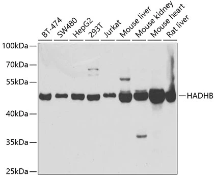
WB analysis of various sample lysates using GTX54152 HADHB antibody. The signal was developed with ECL plus-Enhanced. Dilution : 1:1000 Loading : 25μg per lane

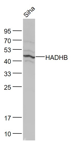
WB analysis of various sample lysates using GTX54152 HADHB antibody. The signal was developed with ECL plus-Enhanced. Dilution : 1:1000 Loading : 25μg per lane
HADHB antibody
GTX54152
ApplicationsImmunoFluorescence, Western Blot, ImmunoCytoChemistry
Product group Antibodies
ReactivityHuman, Mouse, Rat
TargetHADHB
Overview
- SupplierGeneTex
- Product NameHADHB antibody
- Delivery Days Customer7
- Application Supplier NoteWB: 1:500 - 1:2000. ICC/IF: 1:10 - 1:100. *Optimal dilutions/concentrations should be determined by the researcher.Not tested in other applications.
- ApplicationsImmunoFluorescence, Western Blot, ImmunoCytoChemistry
- CertificationResearch Use Only
- ClonalityPolyclonal
- Concentration1 mg/ml
- ConjugateUnconjugated
- Gene ID3032
- Target nameHADHB
- Target descriptionhydroxyacyl-CoA dehydrogenase trifunctional multienzyme complex subunit beta
- Target synonymsECHB, MSTP029, MTPB, MTPD, MTPD2, TP-BETA, trifunctional enzyme subunit beta, mitochondrial, 2-enoyl-Coenzyme A (CoA) hydratase, beta subunit, 3-ketoacyl-Coenzyme A (CoA) thiolase of mitochondrial trifunctional protein, beta subunit, acetyl-CoA acyltransferase, beta-ketothiolase, hydroxyacyl-CoA dehydrogenase/3-ketoacyl-CoA thiolase/enoyl-CoA hydratase (trifunctional protein), beta subunit, hydroxyacyl-Coenzyme A dehydrogenase/3-ketoacyl-Coenzyme A thiolase/enoyl-Coenzyme A hydratase (trifunctional protein), beta subunit
- HostRabbit
- IsotypeIgG
- Protein IDP55084
- Protein NameTrifunctional enzyme subunit beta, mitochondrial
- Scientific DescriptionThis gene encodes the beta subunit of the mitochondrial trifunctional protein, which catalyzes the last three steps of mitochondrial beta-oxidation of long chain fatty acids. The mitochondrial membrane-bound heterocomplex is composed of four alpha and four beta subunits, with the beta subunit catalyzing the 3-ketoacyl-CoA thiolase activity. The encoded protein can also bind RNA and decreases the stability of some mRNAs. The genes of the alpha and beta subunits of the mitochondrial trifunctional protein are located adjacent to each other in the human genome in a head-to-head orientation. Mutations in this gene result in trifunctional protein deficiency. Alternatively spliced transcript variants encoding different isoforms have been described. [provided by RefSeq, Jul 2013]
- ReactivityHuman, Mouse, Rat
- Storage Instruction-20°C or -80°C,2°C to 8°C
- UNSPSC41116161
Datasheet
Related products

Product group Antibodies
Anti-HADHB Antibody Picoband(r)A04776-2-CARRIER-FREE
ApplicationsFlow Cytometry, ImmunoFluorescence, Western Blot, ELISA, ImmunoCytoChemistry, ImmunoHistoChemistry
ReactivityHuman, Mouse, Rat
TargetHADHB
- SizePrice
Product group Antibodies
Anti-HADHB AntibodyA31652
ApplicationsImmunoFluorescence, Western Blot, ImmunoHistoChemistry
ReactivityHuman, Mouse, Rat
- SizePrice
Product group Antibodies
Anti-HADHB Antibody144-05716
ApplicationsImmunoFluorescence, Western Blot
ReactivityHuman, Mouse, Rat
TargetHADHB
- SizePrice
Product group Antibodies
HADHB Polyclonal AntibodyBS-5065R
ApplicationsImmunoFluorescence, Western Blot, ELISA, ImmunoCytoChemistry, ImmunoHistoChemistry, ImmunoHistoChemistry Frozen, ImmunoHistoChemistry Paraffin
ReactivityBovine, Canine, Human, Mouse, Porcine, Rabbit, Rat, Sheep
TargetHADHB
- SizePrice
Product group Antibodies
HADHB Polyclonal AntibodyCAC13256
ApplicationsELISA, ImmunoHistoChemistry
TargetHADHB
- SizePrice
Product group Antibodies
HADHB AntibodyCSB-PA03035A0RB
ApplicationsELISA, ImmunoHistoChemistry
ReactivityHuman
TargetHADHB
- SizePrice
![Various whole cell extracts (30 μg) were separated by 10% SDS-PAGE, and the membrane was blotted with HADHB antibody [N1], N-term (GTX100302) diluted at 1:1000. The HRP-conjugated anti-rabbit IgG antibody (GTX213110-01) was used to detect the primary antibody.](https://www.genetex.com/upload/website/prouct_img/normal/GTX100302/GTX100302_39443_20220121_WB_w_23060100_143.webp)
Product group Antibodies
HADHB antibody [N1], N-termGTX100302
ApplicationsWestern Blot
ReactivityHuman
TargetHADHB
- SizePrice
Product group Antibodies

HADHB AntibodyLS-C482486
ApplicationsImmunoFluorescence, Western Blot, ImmunoCytoChemistry
ReactivityHuman, Mouse, Rat
TargetHADHB
- SizePrice

Product group Antibodies
Anti-HADHB AntibodyHPA037539
ApplicationsWestern Blot, ImmunoHistoChemistry
ReactivityHuman, Mouse, Rat
TargetHADHB
- SizePrice
Product group Antibodies
Anti-HADHB AntibodyCAB5716
ApplicationsImmunoFluorescence, Western Blot, ELISA, ImmunoCytoChemistry
ReactivityHuman
TargetHADHB
- SizePrice