

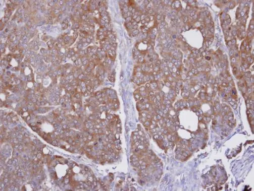
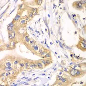
Immunohistochemical analysis of paraffin-embedded NCI-N87 xenograft, using HAGH(GTX105708) antibody at 1:100 dilution.
Antigen Retrieval: Trilogy? (EDTA based, pH 8.0) buffer, 15min
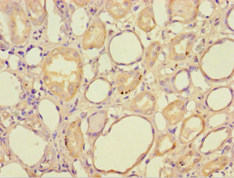
![HAGH antibody [N2C3] detects HAGH protein at mitochondria on mouse kidney by immunohistochemical analysis. Sample: Paraffin-embedded mouse kidney. HAGH antibody [N2C3] (GTX105708) diluted at 1:500.
Antigen Retrieval: Trilogy? (EDTA based, pH 8.0) buffer, 15min](https://www.genetex.com/upload/website/prouct_img/normal/GTX105708/GTX105708_39785_20150116_IHC_M_2_w_23060120_512.webp "HAGH antibody [N2C3] detects HAGH protein at mitochondria on mouse kidney by immunohistochemical analysis. Sample: Paraffin-embedded mouse kidney. HAGH antibody [N2C3] (GTX105708) diluted at 1:500.
Antigen Retrieval: Trilogy? (EDTA based, pH 8.0) buffer, 15min")
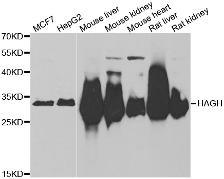
![Mouse tissue extract (50 μg) was separated by 12% SDS-PAGE, and the membrane was blotted with HAGH antibody [N2C3] (GTX105708) diluted at 1:1000. The HRP-conjugated anti-rabbit IgG antibody (GTX213110-01) was used to detect the primary antibody.](https://www.genetex.com/upload/website/prouct_img/normal/GTX105708/GTX105708_39785_20200327_WB_M_liver_w_23060120_270.webp "Mouse tissue extract (50 μg) was separated by 12% SDS-PAGE, and the membrane was blotted with HAGH antibody [N2C3] (GTX105708) diluted at 1:1000. The HRP-conjugated anti-rabbit IgG antibody (GTX213110-01) was used to detect the primary antibody.")
![Whole cell extract (30 μg) was separated by 12% SDS-PAGE, and the membrane was blotted with HAGH antibody [N2C3] (GTX105708) diluted at 1:1000. The HRP-conjugated anti-rabbit IgG antibody (GTX213110-01) was used to detect the primary antibody, and the signal was developed with Trident ECL plus-Enhanced.](https://www.genetex.com/upload/website/prouct_img/normal/GTX105708/GTX105708_45196_20231013_WB_23102401_580.webp "Whole cell extract (30 μg) was separated by 12% SDS-PAGE, and the membrane was blotted with HAGH antibody [N2C3] (GTX105708) diluted at 1:1000. The HRP-conjugated anti-rabbit IgG antibody (GTX213110-01) was used to detect the primary antibody, and the signal was developed with Trident ECL plus-Enhanced.")
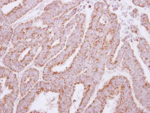
Immunohistochemical analysis of paraffin-embedded NCI-N87 xenograft, using HAGH(GTX105708) antibody at 1:100 dilution.
Antigen Retrieval: Trilogy? (EDTA based, pH 8.0) buffer, 15min
HAGH antibody [N2C3]
GTX105708
ApplicationsWestern Blot, ImmunoHistoChemistry, ImmunoHistoChemistry Paraffin
Product group Antibodies
ReactivityHuman, Mouse
TargetHAGH
Overview
- SupplierGeneTex
- Product NameHAGH antibody [N2C3]
- Delivery Days Customer9
- Application Supplier NoteWB: 1:500-1:3000. IHC-P: 1:100-1:1000. *Optimal dilutions/concentrations should be determined by the researcher.Not tested in other applications.
- ApplicationsWestern Blot, ImmunoHistoChemistry, ImmunoHistoChemistry Paraffin
- CertificationResearch Use Only
- ClonalityPolyclonal
- Concentration1.5 mg/ml
- ConjugateUnconjugated
- Gene ID3029
- Target nameHAGH
- Target descriptionhydroxyacylglutathione hydrolase
- Target synonymsGLO2, GLO2D, GLX2, GLXII, HAGH1, hydroxyacylglutathione hydrolase, mitochondrial, glyoxalase II, hydroxyacylglutathione hydroxylase
- HostRabbit
- IsotypeIgG
- Protein IDQ16775
- Protein NameHydroxyacylglutathione hydrolase, mitochondrial
- Scientific DescriptionThe enzyme encoded by this gene is classified as a thiolesterase and is responsible for the hydrolysis of S-lactoyl-glutathione to reduced glutathione and D-lactate. [provided by RefSeq]
- ReactivityHuman, Mouse
- Storage Instruction-20°C or -80°C,2°C to 8°C
- UNSPSC41116161
Datasheet
Related products
Product group Antibodies
Anti-HAGH AntibodyA31462
ApplicationsWestern Blot, ImmunoHistoChemistry
ReactivityHuman, Mouse, Rat
- SizePrice

Product group Antibodies
Anti-HAGH Antibody Picoband(r)A08943-1-CARRIER-FREE
ApplicationsFlow Cytometry, Western Blot, ELISA, ImmunoHistoChemistry
ReactivityHuman, Mouse, Rat
TargetHAGH
- SizePrice
Product group Antibodies
Anti-HAGH Antibody144-06615
ApplicationsImmunoFluorescence, Western Blot, ImmunoHistoChemistry
ReactivityHuman, Mouse, Rat
TargetHAGH
- SizePrice
Product group Antibodies
HAGH Polyclonal AntibodyBS-15404R
ApplicationsImmunoFluorescence, ELISA, ImmunoCytoChemistry, ImmunoHistoChemistry, ImmunoHistoChemistry Frozen, ImmunoHistoChemistry Paraffin
ReactivityCanine, Equine, Human, Mouse, Rat
- SizePrice
Product group Antibodies
HAGH AntibodyCSB-PA11195A0RB
ApplicationsELISA, ImmunoHistoChemistry
ReactivityHuman
TargetHAGH
- SizePrice
Product group Antibodies

HAGH AntibodyLS-C334831
ApplicationsImmunoFluorescence, Western Blot, ImmunoHistoChemistry
ReactivityHuman
TargetHAGH
- SizePrice

Product group Antibodies
Anti-HAGH AntibodyHPA043041
ApplicationsImmunoHistoChemistry
ReactivityHuman
TargetHAGH
- SizePrice
Product group Antibodies
HAGH antibodyGTX117595
ApplicationsWestern Blot, ImmunoHistoChemistry, ImmunoHistoChemistry Paraffin
ReactivityHuman, Mouse
TargetHAGH
- SizePrice
Product group Antibodies
HAGH antibodyGTX33234
ApplicationsImmunoFluorescence, Western Blot, ImmunoCytoChemistry, ImmunoHistoChemistry, ImmunoHistoChemistry Paraffin
ReactivityHuman, Mouse, Rat
TargetHAGH
- SizePrice
Product group Antibodies
Anti-HAGH AntibodyCAB6615
ApplicationsImmunoFluorescence, Western Blot, ELISA, ImmunoCytoChemistry
ReactivityHuman
TargetHAGH
- SizePrice