

HCG-alpha(HCGa/53), CF405S conjugate, 0.1mg/mL [26628-22-8]
BNC040053
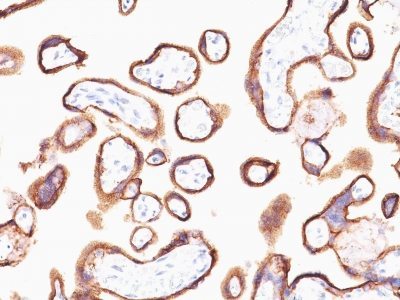
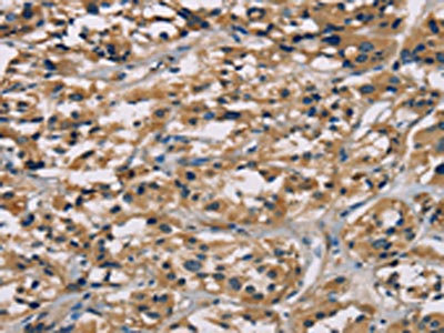
ApplicationsImmunoHistoChemistry, ImmunoHistoChemistry Paraffin
Product group Antibodies
ReactivityBovine, Human, Mouse
TargetCGA
Overview
- SupplierBiotium
- Product NameHCG-alpha(HCGa/53), CF405S conjugate, 0.1mg/mL [26628-22-8]
- Delivery Days Customer9
- ApplicationsImmunoHistoChemistry, ImmunoHistoChemistry Paraffin
- CAS Number26628-22-8
- CertificationResearch Use Only
- ClonalityMonoclonal
- Clone IDHCGa/53
- Concentration0.1 mg/ml
- ConjugateOther Conjugate
- Gene ID1081
- Target nameCGA
- Target descriptionglycoprotein hormones, alpha polypeptide
- Target synonymsCG-ALPHA, FSHA, GPA1, GPHA1, GPHa, HCG, LHA, TSHA, glycoprotein hormones alpha chain, FSH-alpha, LSH-alpha, TSH-alpha, anterior pituitary glycoprotein hormones common subunit alpha, choriogonadotropin alpha chain, chorionic gonadotrophin subunit alpha, chorionic gonadotropin, alpha polypeptide, follicle-stimulating hormone alpha chain, follicle-stimulating hormone alpha subunit, follitropin alpha chain, luteinizing hormone alpha chain, lutropin alpha chain, thyroid-stimulating hormone alpha chain, thyrotropin alpha chain
- HostMouse
- IsotypeIgG1
- Protein IDP01215
- Protein NameGlycoprotein hormones alpha chain
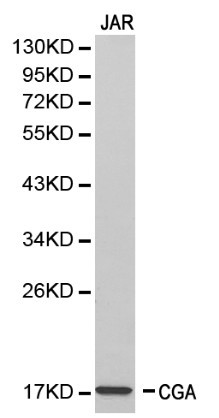
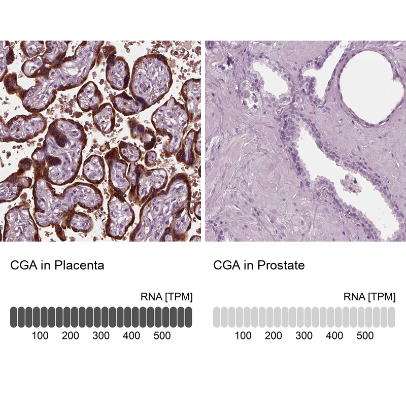
- Scientific DescriptionThis MAb reacts with a protein of ~13 kDa, identified as alpha sub-unit of HCG. HCG is a glycoprotein, which is secreted in large quantities by normal trophoblasts. It is present only in trace amounts in non-pregnant urine and sera but rises sharply during pregnancy. HCG is composed of two non-identical, non-covalently linked polypeptide chains designated as the alpha and beta subunits. The alpha subunit is identical to that of thyroid stimulating hormone (TSH), follicle stimulating hormone (FSH), and luteinizing hormone (LH).Primary antibodies are available purified, or with a selection of fluorescent CF® Dyes and other labels. CF® Dyes offer exceptional brightness and photostability. Note: Conjugates of blue fluorescent dyes like CF®405S and CF®405M are not recommended for detecting low abundance targets, because blue dyes have lower fluorescence and can give higher non-specific background than other dye colors.
- SourceAnimal
- ReactivityBovine, Human, Mouse
- Storage Instruction2°C to 8°C,RT
- UNSPSC41116161
MSDS
Related products
Product group Antibodies
ApplicationsImmunoPrecipitation, Western Blot, ImmunoCytoChemistry, ImmunoHistoChemistry
TargetCGA
- SizePrice

Product group Antibodies
Anti-Chromogranin A/CGA Antibody Picoband(r)A00739-1-CARRIER-FREE
ApplicationsFlow Cytometry, Western Blot, ImmunoHistoChemistry
ReactivityHuman, Mouse, Rat
TargetCGA
- SizePrice
Product group Antibodies
Anti-CGA Antibody144-01239
ApplicationsWestern Blot
ReactivityHuman, Mouse
TargetCGA
- SizePrice
Product group Antibodies

Anti-Thyroid-stimulating hormone [ECACC 88091403]AB01562-1.1-BT
ReactivityHuman
TargetCGA
- SizePrice
Product group Antibodies
Anti-CGA AntibodyA29528
ApplicationsImmunoFluorescence, Western Blot, ImmunoHistoChemistry
ReactivityHuman
- SizePrice
Product group Antibodies
Anti-CGA AntibodyHPA029698
ApplicationsImmunoHistoChemistry
ReactivityHuman
TargetCGA
- SizePrice
Product group Antibodies
CGA AntibodyCSB-PA131153
ApplicationsELISA, ImmunoHistoChemistry
ReactivityHuman
TargetCGA
- SizePrice
Product group Antibodies

CGA / hCG Alpha AntibodyLS-C483329
ApplicationsELISA
ReactivityHuman
TargetCGA
- SizePrice
Product group Antibodies
CGA Recombinant AntibodyBSM-60623R
ApplicationsImmunoFluorescence, Western Blot, ImmunoHistoChemistry, ImmunoHistoChemistry Frozen, ImmunoHistoChemistry Paraffin
ReactivityHuman
TargetCGA
- SizePrice