

Various tissue extracts (50 μg) were separated by 10% SDS-PAGE, and the membrane was blotted with HCK antibody [HL1673] (GTX637272) diluted at 1:1000. The HRP-conjugated anti-rabbit IgG antibody (GTX213110-01) was used to detect the primary antibody.
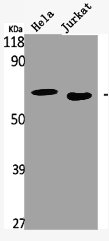
![Various whole cell extracts (30 μg) were separated by 10% SDS-PAGE, and the membrane was blotted with HCK antibody [HL1673] (GTX637272) diluted at 1:1000. The HRP-conjugated anti-rabbit IgG antibody (GTX213110-01) was used to detect the primary antibody.](https://www.genetex.com/upload/website/prouct_img/normal/GTX637272/GTX637272_44809_20220923_WB_22100302_508.webp "Various whole cell extracts (30 μg) were separated by 10% SDS-PAGE, and the membrane was blotted with HCK antibody [HL1673] (GTX637272) diluted at 1:1000. The HRP-conjugated anti-rabbit IgG antibody (GTX213110-01) was used to detect the primary antibody.")
![Whole cell extract (30 μg) was separated by 10% SDS-PAGE, and the membrane was blotted with HCK antibody [HL1673] (GTX637272) diluted at 1:1000. The HRP-conjugated anti-rabbit IgG antibody (GTX213110-01) was used to detect the primary antibody.](https://www.genetex.com/upload/website/prouct_img/normal/GTX637272/GTX637272_44809_20220930_WB_22101319_817.webp "Whole cell extract (30 μg) was separated by 10% SDS-PAGE, and the membrane was blotted with HCK antibody [HL1673] (GTX637272) diluted at 1:1000. The HRP-conjugated anti-rabbit IgG antibody (GTX213110-01) was used to detect the primary antibody.")
![HCK antibody [HL1673] detects HCK protein at cell membrane and cytoplasm by immunohistochemical analysis. Sample: Paraffin-embedded human hepatocellular carcinoma. HCK stained by HCK antibody [HL1673] (GTX637272) diluted at 1:100. Antigen Retrieval: Citrate buffer, pH 6.0, 15 min](https://www.genetex.com/upload/website/prouct_img/normal/GTX637272/GTX637272_44809_20221223_IHC-P_22122722_724.webp "HCK antibody [HL1673] detects HCK protein at cell membrane and cytoplasm by immunohistochemical analysis. Sample: Paraffin-embedded human hepatocellular carcinoma. HCK stained by HCK antibody [HL1673] (GTX637272) diluted at 1:100. Antigen Retrieval: Citrate buffer, pH 6.0, 15 min")
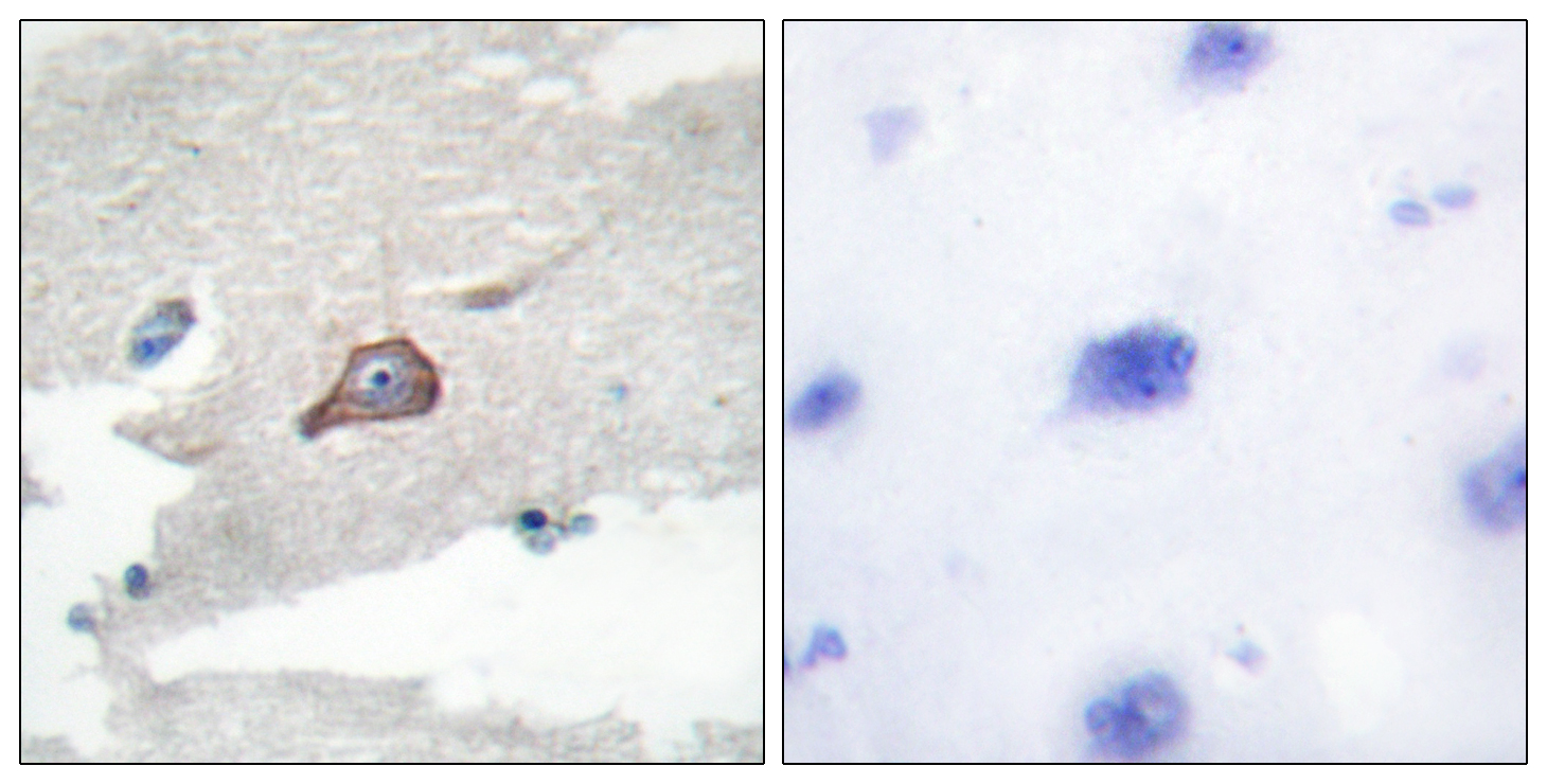
![HCK antibody [HL1673] detects HCK protein at cell membrane and cytoplasm by immunohistochemical analysis. Sample: Paraffin-embedded mouse tissues. HCK stained by HCK antibody [HL1673] (GTX637272) diluted at 1:100. Antigen Retrieval: Citrate buffer, pH 6.0, 15 min](https://www.genetex.com/upload/website/prouct_img/normal/GTX637272/GTX637272_44809_20221223_IHC-P_M_22122722_258.webp "HCK antibody [HL1673] detects HCK protein at cell membrane and cytoplasm by immunohistochemical analysis. Sample: Paraffin-embedded mouse tissues. HCK stained by HCK antibody [HL1673] (GTX637272) diluted at 1:100. Antigen Retrieval: Citrate buffer, pH 6.0, 15 min")
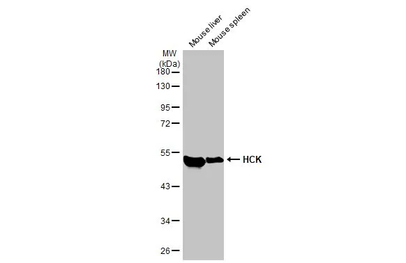
Various tissue extracts (50 μg) were separated by 10% SDS-PAGE, and the membrane was blotted with HCK antibody [HL1673] (GTX637272) diluted at 1:1000. The HRP-conjugated anti-rabbit IgG antibody (GTX213110-01) was used to detect the primary antibody.
HCK antibody [HL1673]
GTX637272
ApplicationsWestern Blot, ImmunoHistoChemistry, ImmunoHistoChemistry Paraffin
Product group Antibodies
ReactivityHuman, Mouse
TargetHCK
Overview
- SupplierGeneTex
- Product NameHCK antibody [HL1673]
- Delivery Days Customer9
- Application Supplier NoteWB: 1:500-1:3000. *Optimal dilutions/concentrations should be determined by the researcher.Not tested in other applications.
- ApplicationsWestern Blot, ImmunoHistoChemistry, ImmunoHistoChemistry Paraffin
- CertificationResearch Use Only
- ClonalityMonoclonal
- Clone IDHL1673
- Concentration1 mg/ml
- ConjugateUnconjugated
- Gene ID3055
- Target nameHCK
- Target descriptionHCK proto-oncogene, Src family tyrosine kinase
- Target synonymsAIPCV, JTK9, p59Hck, p61Hck, tyrosine-protein kinase HCK, hematopoietic cell kinase, hemopoietic cell kinase, p59-HCK/p60-HCK
- HostRabbit
- IsotypeIgG
- Protein IDP08631
- Protein NameTyrosine-protein kinase HCK
- Scientific DescriptionThe protein encoded by this gene is a member of the Src family of tyrosine kinases. This protein is primarily hemopoietic, particularly in cells of the myeloid and B-lymphoid lineages. It may help couple the Fc receptor to the activation of the respiratory burst. In addition, it may play a role in neutrophil migration and in the degranulation of neutrophils. Multiple isoforms with different subcellular distributions are produced due to both alternative splicing and the use of alternative translation initiation codons, including a non-AUG (CUG) codon. [provided by RefSeq, Feb 2010]
- ReactivityHuman, Mouse
- Storage Instruction-20°C or -80°C,2°C to 8°C
- UNSPSC41116161
Datasheet
Related products
Product group Antibodies
Anti-HCK AntibodyA96452
ApplicationsImmunoFluorescence, ELISA, ImmunoHistoChemistry
ReactivityHuman, Mouse, Rat
- SizePrice
Product group Antibodies
Anti-HCK Antibody144-60699
ApplicationsWestern Blot
ReactivityHuman, Mouse, Rat
TargetHCK
- SizePrice
Product group Antibodies
HCK AntibodyCSB-PA002872
ApplicationsImmunoFluorescence, Western Blot, ELISA, ImmunoHistoChemistry
ReactivityHuman, Mouse, Rat
TargetHCK
- SizePrice
Product group Antibodies
ApplicationsFlow Cytometry
TargetHCK
- SizePrice
Product group Antibodies
Hck Polyclonal AntibodyBS-1438R
ApplicationsImmunoFluorescence, Western Blot, ELISA, ImmunoCytoChemistry, ImmunoHistoChemistry, ImmunoHistoChemistry Frozen, ImmunoHistoChemistry Paraffin
ReactivityBovine, Chicken, Equine, Human, Mouse, Rabbit, Rat, Sheep
TargetHCK
- SizePrice
Product group Antibodies

HCK AntibodyLS-C404038
ApplicationsWestern Blot, ELISA
ReactivityHuman, Mouse, Rat
TargetHCK
- SizePrice
![IHC-P analysis of human colon cancer (left) and ancreas cancer (right) using GTX83203 HCK antibody [3D12E10].](https://www.genetex.com/upload/website/prouct_img/normal/GTX83203/GTX83203_20170912_IHC-P_w_23061322_902.webp)
Product group Antibodies
HCK antibody [3D12E10]GTX83203
ApplicationsWestern Blot, ELISA, ImmunoHistoChemistry, ImmunoHistoChemistry Paraffin
ReactivityHuman
TargetHCK
- SizePrice
Product group Antibodies
HCK (phospho Tyr521) antibodyGTX55374
ApplicationsWestern Blot
ReactivityHuman
TargetHCK
- SizePrice
Product group Antibodies
HCK antibodyGTX32645
ApplicationsImmunoFluorescence, Western Blot, ImmunoCytoChemistry
ReactivityHuman
TargetHCK
- SizePrice