

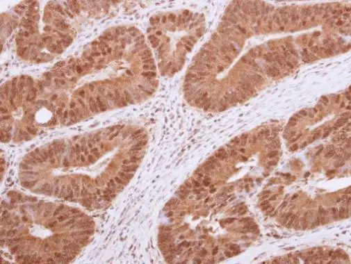
Immunohistochemical analysis of paraffin-embedded human colon carcinoma, using HDAC4(GTX110231) antibody at 1:250 dilution.
Antigen Retrieval: Trilogy? (EDTA based, pH 8.0) buffer, 15min
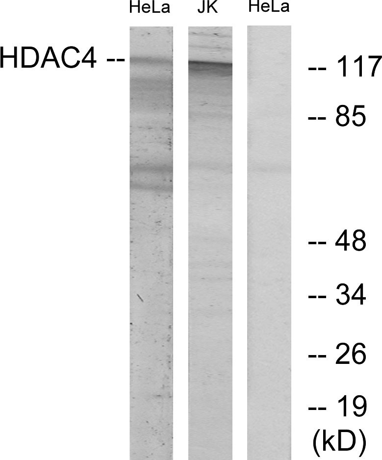
![Various whole cell extracts (30 μg) were separated by 7.5% SDS-PAGE, and the membrane was blotted with HDAC4 antibody [N3C1], Internal (GTX110231) diluted at 1:1000. The HRP-conjugated anti-rabbit IgG antibody (GTX213110-01) was used to detect the primary antibody.](https://www.genetex.com/upload/website/prouct_img/normal/GTX110231/GTX110231_42795_20181221_WB_M_w_23060500_150.webp "Various whole cell extracts (30 μg) were separated by 7.5% SDS-PAGE, and the membrane was blotted with HDAC4 antibody [N3C1], Internal (GTX110231) diluted at 1:1000. The HRP-conjugated anti-rabbit IgG antibody (GTX213110-01) was used to detect the primary antibody.")
 of paraformaldehyde-fixed HeLa, using HDAC4(GTX110231) antibody (Green) at 1:500 dilution. Alpha-tubulin filaments were labeled with GTX11304 (Red) at 1:2000.")
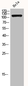
![Various whole cell extracts (30 μg) were separated by 5% SDS-PAGE, and the membrane was blotted with HDAC4 antibody [N3C1], Internal (GTX110231) diluted at 1:1000. The HRP-conjugated anti-rabbit IgG antibody (GTX213110-01) was used to detect the primary antibody.](https://www.genetex.com/upload/website/prouct_img/normal/GTX110231/GTX110231_43754_20191101_WB_w_23060500_277.webp "Various whole cell extracts (30 μg) were separated by 5% SDS-PAGE, and the membrane was blotted with HDAC4 antibody [N3C1], Internal (GTX110231) diluted at 1:1000. The HRP-conjugated anti-rabbit IgG antibody (GTX213110-01) was used to detect the primary antibody.")
![Various whole cell extracts (30 μg) were separated by 7.5% SDS-PAGE, and the membrane was blotted with HDAC4 antibody [N3C1], Internal (GTX110231) diluted at 1:1000. The HRP-conjugated anti-rabbit IgG antibody (GTX213110-01) was used to detect the primary antibody.](https://www.genetex.com/upload/website/prouct_img/normal/GTX110231/GTX110231_42795_20181221_WB_R_w_23060500_137.webp "Various whole cell extracts (30 μg) were separated by 7.5% SDS-PAGE, and the membrane was blotted with HDAC4 antibody [N3C1], Internal (GTX110231) diluted at 1:1000. The HRP-conjugated anti-rabbit IgG antibody (GTX213110-01) was used to detect the primary antibody.")
![Non-transfected (–) and transfected (+) 293T whole cell extracts (60 μg) were separated by 7.5% SDS-PAGE, and the membrane was blotted with HDAC4 antibody [N3C1], Internal (GTX110231) diluted at 1:1000. The HRP-conjugated anti-rabbit IgG antibody (GTX213110-01) was used to detect the primary antibody.](https://www.genetex.com/upload/website/prouct_img/normal/GTX110231/GTX110231_42795_20181026_WB_shRNA_watermark_w_23060500_628.webp "Non-transfected (–) and transfected (+) 293T whole cell extracts (60 μg) were separated by 7.5% SDS-PAGE, and the membrane was blotted with HDAC4 antibody [N3C1], Internal (GTX110231) diluted at 1:1000. The HRP-conjugated anti-rabbit IgG antibody (GTX213110-01) was used to detect the primary antibody.")
![HDAC4 antibody [N3C1], Internal immunoprecipitates HDAC4 protein in IP experiments. IP samples: Jurkat whole cell extract A. Control with 4 μg of preimmune Rabbit IgG B. Immunoprecipitation of HDAC4 protein by 4 μg HDAC4 antibody [N3C1], Internal (GTX110231) 5 % SDS-PAGE The immunoprecipitated HDAC4 protein was detected by HDAC4 antibody [N3C1], Internal (GTX110231) diluted at 1:500. [EasyBlot anti-rabbit IgG (GTX221666-01) was used as a secondary reagent]](https://www.genetex.com/upload/website/prouct_img/normal/GTX110231/GTX110231_40478_IP_w_23060500_315.webp "HDAC4 antibody [N3C1], Internal immunoprecipitates HDAC4 protein in IP experiments. IP samples: Jurkat whole cell extract A. Control with 4 μg of preimmune Rabbit IgG B. Immunoprecipitation of HDAC4 protein by 4 μg HDAC4 antibody [N3C1], Internal (GTX110231) 5 % SDS-PAGE The immunoprecipitated HDAC4 protein was detected by HDAC4 antibody [N3C1], Internal (GTX110231) diluted at 1:500. [EasyBlot anti-rabbit IgG (GTX221666-01) was used as a secondary reagent]")
Immunohistochemical analysis of paraffin-embedded human colon carcinoma, using HDAC4(GTX110231) antibody at 1:250 dilution.
Antigen Retrieval: Trilogy? (EDTA based, pH 8.0) buffer, 15min
HDAC4 antibody [N3C1], Internal
GTX110231
ApplicationsImmunoFluorescence, ImmunoPrecipitation, Western Blot, ImmunoCytoChemistry, ImmunoHistoChemistry, ImmunoHistoChemistry Frozen, ImmunoHistoChemistry Paraffin
Product group Antibodies
ReactivityHuman, Mouse, Rat
TargetHDAC4
Overview
- SupplierGeneTex
- Product NameHDAC4 antibody [N3C1], Internal
- Delivery Days Customer9
- Application Supplier NoteWB: 1:500-1:3000. ICC/IF: 1:100-1:1000. IHC-P: 1:100-1:1000. IP: 1:100-1:500. *Optimal dilutions/concentrations should be determined by the researcher.Not tested in other applications.
- ApplicationsImmunoFluorescence, ImmunoPrecipitation, Western Blot, ImmunoCytoChemistry, ImmunoHistoChemistry, ImmunoHistoChemistry Frozen, ImmunoHistoChemistry Paraffin
- CertificationResearch Use Only
- ClonalityPolyclonal
- Concentration1.72 mg/ml
- ConjugateUnconjugated
- Gene ID9759
- Target nameHDAC4
- Target descriptionhistone deacetylase 4
- Target synonymsAHO3, BDMR, HA6116, HD4, HDAC-4, HDAC-A, HDACA, NEDCHF, NEDCHID, histone deacetylase 4, histone deacetylase A
- HostRabbit
- IsotypeIgG
- Protein IDP56524
- Protein NameHistone deacetylase 4
- Scientific DescriptionHistones play a critical role in transcriptional regulation, cell cycle progression, and developmental events. Histone acetylation/deacetylation alters chromosome structure and affects transcription factor access to DNA. The protein encoded by this gene belongs to class II of the histone deacetylase/acuc/apha family. It possesses histone deacetylase activity and represses transcription when tethered to a promoter. This protein does not bind DNA directly, but through transcription factors MEF2C and MEF2D. It seems to interact in a multiprotein complex with RbAp48 and HDAC3. [provided by RefSeq]
- ReactivityHuman, Mouse, Rat
- Storage Instruction-20°C or -80°C,2°C to 8°C
- UNSPSC41116161
Datasheet
Related products
Product group Antibodies
Anti-HDAC4 AntibodyA97494
ApplicationsWestern Blot, ELISA
ReactivityHuman, Mouse, Rat
- SizePrice
Product group Antibodies

Anti-HDAC4 [RAB-C320]Ab01781-1.1
ApplicationsImmunoPrecipitation
ReactivityHuman
TargetHDAC4
- SizePrice

Product group Antibodies
Anti-HDAC4 Antibody Picoband(r)A00971-1-CARRIER-FREE
ApplicationsFlow Cytometry, ImmunoFluorescence, Western Blot, ImmunoCytoChemistry, ImmunoHistoChemistry
ReactivityHuman, Mouse, Rat
TargetHDAC4
- SizePrice
Product group Antibodies
Anti-HDAC4 Antibody144-00179
ApplicationsWestern Blot, ImmunoHistoChemistry
ReactivityHuman, Mouse, Rat
TargetHDAC4
- SizePrice
Product group Antibodies

HDAC4 AntibodyLS-C746719
ApplicationsWestern Blot, ImmunoHistoChemistry
ReactivityHuman, Mouse, Rat
TargetHDAC4
- SizePrice
Product group Antibodies
ApplicationsImmunoFluorescence, Western Blot, ImmunoHistoChemistry, ImmunoHistoChemistry Frozen, ImmunoHistoChemistry Paraffin
ReactivityHuman
TargetHDAC4
- SizePrice
Product group Antibodies
HDAC4 AntibodyCSB-PA002881
ApplicationsWestern Blot, ELISA
ReactivityHuman, Mouse, Rat
TargetHDAC4
- SizePrice
Product group Antibodies
ApplicationsImmunoPrecipitation, Western Blot, ImmunoCytoChemistry, ImmunoHistoChemistry
TargetHDAC4
- SizePrice
![WB analysis of HeLa (1), Jurkat (2) cell lysate using GTX83229 HDAC4 antibody [7B2].](https://www.genetex.com/upload/website/prouct_img/normal/GTX83229/GTX83229_20170912_WB_w_23061322_126.webp)
Product group Antibodies
HDAC4 antibody [7B2]GTX83229
ApplicationsWestern Blot, ELISA
ReactivityHuman
TargetHDAC4
- SizePrice

Product group Antibodies
Anti-HDAC4 AntibodyHPA048723
ApplicationsImmunoCytoChemistry
ReactivityHuman
TargetHDAC4
- SizePrice