

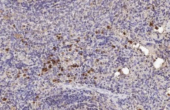
Hemoglobin epsilon antibody [HL2457] detects secreted Hemoglobin epsilon protein by immunohistochemical analysis. Sample: Paraffin-embedded rat spleen. Hemoglobin epsilon stained by Hemoglobin epsilon antibody [HL2457] (GTX638775) diluted at 1:100. Antigen Retrieval: Citrate buffer, pH 6.0, 15 min
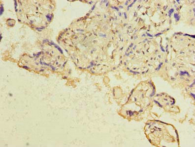
![Hemoglobin epsilon antibody [HL2457] detects secreted Hemoglobin epsilon protein by immunohistochemical analysis. Sample: Paraffin-embedded mouse spleen. Hemoglobin epsilon stained by Hemoglobin epsilon antibody [HL2457] (GTX638775) diluted at 1:100. Antigen Retrieval: Citrate buffer, pH 6.0, 15 min](https://www.genetex.com/upload/website/prouct_img/normal/GTX638775/GTX638775_T-45089_20230721_IHC-P_M_23073119_222.webp "Hemoglobin epsilon antibody [HL2457] detects secreted Hemoglobin epsilon protein by immunohistochemical analysis. Sample: Paraffin-embedded mouse spleen. Hemoglobin epsilon stained by Hemoglobin epsilon antibody [HL2457] (GTX638775) diluted at 1:100. Antigen Retrieval: Citrate buffer, pH 6.0, 15 min")
![Hemoglobin epsilon antibody [HL2457] detects Hemoglobin epsilon protein at cell membrane and cytoplasm by immunofluorescent analysis. Sample: K562 cells were fixed in 4% paraformaldehyde at RT for 15 min. Green: Hemoglobin epsilon stained by Hemoglobin epsilon antibody [HL2457] (GTX638775) diluted at 1:500. Blue: Fluoroshield with DAPI (GTX30920).](https://www.genetex.com/upload/website/prouct_img/normal/GTX638775/GTX638775_T-45089_20230825_ICC_IF_23090619_621.webp "Hemoglobin epsilon antibody [HL2457] detects Hemoglobin epsilon protein at cell membrane and cytoplasm by immunofluorescent analysis. Sample: K562 cells were fixed in 4% paraformaldehyde at RT for 15 min. Green: Hemoglobin epsilon stained by Hemoglobin epsilon antibody [HL2457] (GTX638775) diluted at 1:500. Blue: Fluoroshield with DAPI (GTX30920).")
![Various whole cell extracts (30 μg) were separated by 15% SDS-PAGE, and the membrane was blotted with Hemoglobin epsilon antibody [HL2457] (GTX638775) diluted at 1:1000. The HRP-conjugated anti-rabbit IgG antibody (GTX213110-01) was used to detect the primary antibody. Corresponding RNA expression data for the same cell lines are based on Human Protein Atlas program.](https://www.genetex.com/upload/website/prouct_img/normal/GTX638775/GTX638775_45159_20230908_WB_TPM_watermark_23091319_397.webp "Various whole cell extracts (30 μg) were separated by 15% SDS-PAGE, and the membrane was blotted with Hemoglobin epsilon antibody [HL2457] (GTX638775) diluted at 1:1000. The HRP-conjugated anti-rabbit IgG antibody (GTX213110-01) was used to detect the primary antibody. Corresponding RNA expression data for the same cell lines are based on Human Protein Atlas program.")
Hemoglobin epsilon antibody [HL2457] detects secreted Hemoglobin epsilon protein by immunohistochemical analysis. Sample: Paraffin-embedded rat spleen. Hemoglobin epsilon stained by Hemoglobin epsilon antibody [HL2457] (GTX638775) diluted at 1:100. Antigen Retrieval: Citrate buffer, pH 6.0, 15 min
Hemoglobin epsilon antibody [HL2457]
GTX638775
ApplicationsImmunoFluorescence, Western Blot, ImmunoCytoChemistry, ImmunoHistoChemistry, ImmunoHistoChemistry Paraffin
Product group Antibodies
ReactivityHuman, Mouse, Rat
TargetHBE1
Overview
- SupplierGeneTex
- Product NameHemoglobin epsilon antibody [HL2457]
- Delivery Days Customer9
- Application Supplier NoteWB: 1:500-1:3000. *Optimal dilutions/concentrations should be determined by the researcher.Not tested in other applications.
- ApplicationsImmunoFluorescence, Western Blot, ImmunoCytoChemistry, ImmunoHistoChemistry, ImmunoHistoChemistry Paraffin
- CertificationResearch Use Only
- ClonalityMonoclonal
- Clone IDHL2457
- Concentration1 mg/ml
- ConjugateUnconjugated
- Gene ID3046
- Target nameHBE1
- Target descriptionhemoglobin subunit epsilon 1
- Target synonymsHBE, hemoglobin subunit epsilon, epsilon globin, hemoglobin epsilon chain, hemoglobin, epsilon 1
- HostRabbit
- IsotypeIgG
- Protein IDP02100
- Protein NameHemoglobin subunit epsilon
- Scientific DescriptionThe epsilon globin gene (HBE) is normally expressed in the embryonic yolk sac: two epsilon chains together with two zeta chains (an alpha-like globin) constitute the embryonic hemoglobin Hb Gower I; two epsilon chains together with two alpha chains form the embryonic Hb Gower II. Both of these embryonic hemoglobins are normally supplanted by fetal, and later, adult hemoglobin. The five beta-like globin genes are found within a 45 kb cluster on chromosome 11 in the following order: 5-epsilon - G-gamma - A-gamma - delta - beta-3 [provided by RefSeq, Jul 2008]
- ReactivityHuman, Mouse, Rat
- Storage Instruction-20°C or -80°C,2°C to 8°C
- UNSPSC41116161
Datasheet
Related products

Product group Antibodies
Anti-HBE1 Antibody(Center)A08093-1
ApplicationsFlow Cytometry, Western Blot, ImmunoHistoChemistry, ImmunoHistoChemistry Paraffin
ReactivityHuman, Rabbit
TargetHBE1
- SizePrice
Product group Antibodies

HBE1 / Hemoglobin Epsilon 1 AntibodyLS-C830667
ApplicationsWestern Blot, ELISA, ImmunoHistoChemistry
ReactivityHuman
TargetHBE1
- SizePrice
Product group Antibodies
HBE1 AntibodyCSB-PA010153LA01HU
ApplicationsELISA, ImmunoHistoChemistry
ReactivityHuman
TargetHBE1
- SizePrice
Product group Antibodies
ApplicationsImmunoPrecipitation, Western Blot, ImmunoCytoChemistry, ImmunoHistoChemistry
TargetHBE1
- SizePrice
![Various tissue extracts (50 μg) were separated by 15% SDS-PAGE, and the membrane was blotted with Hemoglobin epsilon antibody [HL2458] (GTX638776) diluted at 1:5000. The HRP-conjugated anti-rabbit IgG antibody (GTX213110-01) was used to detect the primary antibody.](https://www.genetex.com/upload/website/prouct_img/normal/GTX638776/GTX638776_T-45089_20230714_WB_M_R_23071822_352.webp)
Product group Antibodies
Hemoglobin epsilon antibody [HL2458]GTX638776
ApplicationsWestern Blot, ImmunoHistoChemistry, ImmunoHistoChemistry Paraffin
ReactivityHuman, Mouse, Rat
TargetHBE1
- SizePrice
![Untreated (–) and treated (+) mouse tissue extracts (30 μg) were separated by 15% SDS-PAGE, and the membrane was blotted with Hemoglobin epsilon antibody [N1N2], N-term (GTX108396) diluted at 1:500. (CFA: Complete Freunds adjuvant)](https://www.genetex.com/upload/website/prouct_img/normal/GTX108396/GTX108396_39848_20160331_WB_M_CFA_w_23060120_798.webp)
Product group Antibodies
ApplicationsWestern Blot, ImmunoHistoChemistry, ImmunoHistoChemistry Paraffin
ReactivityHuman, Mouse, Rat
TargetHBE1
- SizePrice