

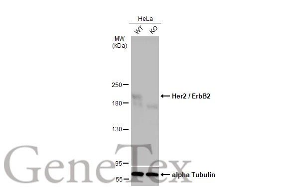
Wild-type (WT) and Her2 / ErbB2 knockout (KO) HeLa cell extracts (30 μg) were separated by 5% SDS-PAGE, and the membrane was blotted with Her2 / ErbB2 antibody (GTX100509) diluted at 1:5000. The HRP-conjugated anti-rabbit IgG antibody (GTX213110-01) was used to detect the primary antibody, and the signal was developed with Trident femto Western HRP Substrate.
![Various whole cell extracts (30 μg) were separated by 5% SDS-PAGE, and the membrane was blotted with Her2 / ErbB2 antibody [C2C3], C-term (GTX100509) diluted at 1:2000. The HRP-conjugated anti-rabbit IgG antibody (GTX213110-01) was used to detect the primary antibody.](https://www.genetex.com/upload/website/prouct_img/normal/GTX100509/GTX100509_41332_20170818_WB_w_23060100_664.webp "Various whole cell extracts (30 μg) were separated by 5% SDS-PAGE, and the membrane was blotted with Her2 / ErbB2 antibody [C2C3], C-term (GTX100509) diluted at 1:2000. The HRP-conjugated anti-rabbit IgG antibody (GTX213110-01) was used to detect the primary antibody.")
 and transfected (+) 293T whole cell extracts were separated by 5% SDS-PAGE, and the membrane was blotted with Her2 / ErbB2 antibody (GTX100509) diluted at 1:7000. The HRP-conjugated anti-rabbit IgG antibody (GTX213110-01) was used to detect the primary antibody.")
![Various whole cell extracts (30 μg) were separated by 5% SDS-PAGE, and the membrane was blotted with Her2 / ErbB2 antibody [C2C3], C-term (GTX100509) diluted at 1:2000. The HRP-conjugated anti-rabbit IgG antibody (GTX213110-01) was used to detect the primary antibody.](https://www.genetex.com/upload/website/prouct_img/normal/GTX100509/GTX100509_41332_20170818_WB_2_w_23060100_351.webp "Various whole cell extracts (30 μg) were separated by 5% SDS-PAGE, and the membrane was blotted with Her2 / ErbB2 antibody [C2C3], C-term (GTX100509) diluted at 1:2000. The HRP-conjugated anti-rabbit IgG antibody (GTX213110-01) was used to detect the primary antibody.")
 diluted at 1:500.")
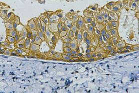
![Her2 / ErbB2 antibody [C2C3], C-term detects Her2 / ErbB2 protein at cell membrane in human breast carcinoma by immunohistochemical analysis. Sample: Strong positive (++), low positive (+) and negative tissue slides cores assessed using Quantitative Digital Pathology. Her2 / ErbB2 antibody [C2C3], C-term (GTX100509) diluted at 1:500, and competitor's antibody (CST#4290) diluted at 1:100.
Antigen Retrieval: Trilogy? (EDTA based, pH 8.0) buffer, 15min](https://www.genetex.com/upload/website/prouct_img/normal/GTX100509/GTX100509_41332_20170817_IHC-P_H_w_23060100_926.webp "Her2 / ErbB2 antibody [C2C3], C-term detects Her2 / ErbB2 protein at cell membrane in human breast carcinoma by immunohistochemical analysis. Sample: Strong positive (++), low positive (+) and negative tissue slides cores assessed using Quantitative Digital Pathology. Her2 / ErbB2 antibody [C2C3], C-term (GTX100509) diluted at 1:500, and competitor's antibody (CST#4290) diluted at 1:100.
Antigen Retrieval: Trilogy? (EDTA based, pH 8.0) buffer, 15min")
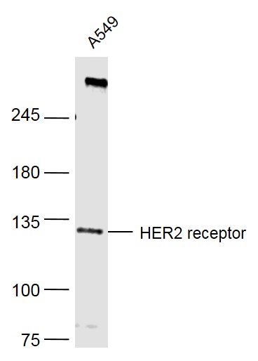
![Whole cell extract (30 μg) was separated by 5% SDS-PAGE, and the membrane was blotted with Her2 / ErbB2 antibody [C2C3], C-term (GTX100509) diluted at 1:10000.](https://www.genetex.com/upload/website/prouct_img/normal/GTX100509/GTX100509_41332_20160414_WB_w_23060100_632.webp "Whole cell extract (30 μg) was separated by 5% SDS-PAGE, and the membrane was blotted with Her2 / ErbB2 antibody [C2C3], C-term (GTX100509) diluted at 1:10000.")
 diluted at 1:50.")
 were separated by 7.5% SDS-PAGE, and the membrane was blotted with Her2 / ErbB2 antibody (GTX100509) diluted at 1:1000. The HRP-conjugated anti-rabbit IgG antibody (GTX213110-01) was used to detect the primary antibody. Corresponding RNA expression data for the same cell lines are based on Human Protein Atlas program.")
Wild-type (WT) and Her2 / ErbB2 knockout (KO) HeLa cell extracts (30 μg) were separated by 5% SDS-PAGE, and the membrane was blotted with Her2 / ErbB2 antibody (GTX100509) diluted at 1:5000. The HRP-conjugated anti-rabbit IgG antibody (GTX213110-01) was used to detect the primary antibody, and the signal was developed with Trident femto Western HRP Substrate.
Her2 / ErbB2 antibody
GTX100509
ApplicationsWestern Blot, ImmunoHistoChemistry, ImmunoHistoChemistry Paraffin
Product group Antibodies
ReactivityHuman
TargetERBB2
Overview
- SupplierGeneTex
- Product NameHer2 / ErbB2 antibody
- Delivery Days Customer9
- Application Supplier NoteWB: 1:500-1:20000. IHC-P: 1:100-1:1000. *Optimal dilutions/concentrations should be determined by the researcher.Not tested in other applications.
- ApplicationsWestern Blot, ImmunoHistoChemistry, ImmunoHistoChemistry Paraffin
- CertificationResearch Use Only
- ClonalityPolyclonal
- Concentration0.07 mg/ml
- ConjugateUnconjugated
- Gene ID2064
- Target nameERBB2
- Target descriptionerb-b2 receptor tyrosine kinase 2
- Target synonymsCD340, HER-2, HER-2/neu, HER2, MLN 19, MLN-19, NEU, NGL, TKR1, VSCN2, c-ERB-2, c-ERB2, p185(erbB2), receptor tyrosine-protein kinase erbB-2, c-erb B2/neu protein, herstatin, human epidermal growth factor receptor 2, metastatic lymph node gene 19 protein, neuro/glioblastoma derived oncogene homolog, neuroblastoma/glioblastoma derived oncogene homolog, proto-oncogene Neu, proto-oncogene c-ErbB-2, tyrosine kinase-type cell surface receptor HER2, v-erb-b2 avian erythroblastic leukemia viral oncogene homolog 2, v-erb-b2 avian erythroblastic leukemia viral oncoprotein 2, v-erb-b2 erythroblastic leukemia viral oncogene homolog 2, neuro/glioblastoma derived oncogene homolog
- HostRabbit
- IsotypeIgG
- Protein IDP04626
- Protein NameReceptor tyrosine-protein kinase erbB-2
- Scientific DescriptionThis gene encodes a member of the epidermal growth factor (EGF) receptor family of receptor tyrosine kinases. This protein has no ligand binding domain of its own and therefore cannot bind growth factors. However, it does bind tightly to other ligand-bound EGF receptor family members to form a heterodimer, stabilizing ligand binding and enhancing kinase-mediated activation of downstream signalling pathways, such as those involving mitogen-activated protein kinase and phosphatidylinositol-3 kinase. Allelic variations at amino acid positions 654 and 655 of isoform a (positions 624 and 625 of isoform b) have been reported, with the most common allele, Ile654/Ile655, shown here. Amplification and/or overexpression of this gene has been reported in numerous cancers, including breast and ovarian tumors. Alternative splicing results in several additional transcript variants, some encoding different isoforms and others that have not been fully characterized. [provided by RefSeq]
- ReactivityHuman
- Storage Instruction-20°C or -80°C,2°C to 8°C
- UNSPSC41116161
Datasheet
Related products
Product group Antibodies
Anti-HER2 AntibodyA95367
ApplicationsImmunoFluorescence, Western Blot, ELISA, ImmunoHistoChemistry
ReactivityHuman, Mouse, Rat
- SizePrice
Product group Antibodies

Anti-erbB-2 (Her-2/neu) [4D5-8 (trastuzumab)]Ab00103-10.0
ApplicationsFlow Cytometry, ELISA, ImmunoHistoChemistry
ReactivityHuman
TargetERBB2
- SizePrice
Product group Antibodies
Anti-Her2/Neu Antibody118-10015
ApplicationsWestern Blot, ELISA, ImmunoHistoChemistry
ReactivityHuman
- SizePrice

Product group Antibodies
Anti-HER2 AntibodyAMAB90627
ApplicationsWestern Blot, ImmunoHistoChemistry
ReactivityHuman
TargetERBB2
- SizePrice
Product group Antibodies
HER2 (1A4) Monoclonal AntibodyBSM-2156M
ApplicationsImmunoFluorescence, Western Blot, ImmunoCytoChemistry, ImmunoHistoChemistry, ImmunoHistoChemistry Frozen, ImmunoHistoChemistry Paraffin
ReactivityHuman
TargetERBB2
- SizePrice

Product group Antibodies
Anti-ErbB 2/ERBB2 Antibody Picoband(r)A00010-2-CARRIER-FREE
ApplicationsWestern Blot, ImmunoHistoChemistry
ReactivityHuman
TargetERBB2
- SizePrice
Product group Antibodies
ERBB2 Monoclonal AntibodyCSB-MA000200
ApplicationsWestern Blot, ELISA, ImmunoHistoChemistry
ReactivityHuman, Mouse, Rat
TargetERBB2
- SizePrice
Product group Antibodies

Goat anti-ERBB2 / HER2EB11748
ApplicationsELISA, ImmunoHistoChemistry
ReactivityHuman
TargetERBB2
- SizePrice
Product group Antibodies
Erbb2 Polyclonal AntibodyCAC09113
ApplicationsELISA, ImmunoHistoChemistry
TargetERBB2
- SizePrice