

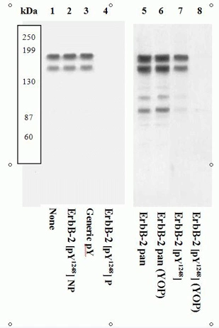
Extracts of SK-BR-3 cells were resolved by SDS-PAGE on a 10% Tris-glycine gel and transferred to PVDF. The membrane was either not treated (1-5, 7) or treated (6, 8) with YOP phosphatase, blocked with a 5% BSA-TBST buffer overnight at 4oC, then incubated with the ErbB-2 [pY1248] antibody for two hours at room temperature in a 3% BSA-TBST buffer, following prior incubation with: no peptide (1, 5-8), the non-phosphopeptide corresponding to the phosphopeptide immunogen (2), a generic phosphotyrosine-containing peptide (3), or the phosphopeptide immunogen (4). After washing, the membrane was incubated with goat F(ab)2 anti-rabbit IgG alkaline phosphatase and signals were detected.The data show that only the phosphopeptide corresponding to ErbB-2 [pY1248] blocks the antibody signal, demonstrating the specificity of the antibody. The data also show that phosphatase stripping eliminates the signal, further verifying that the antibody is phospho-specific.
Extracts of SK-BR-3 cells were resolved by SDS-PAGE on a 10% Tris-glycine gel and transferred to PVDF. The membrane was either not treated (1-5, 7) or treated (6, 8) with YOP phosphatase, blocked with a 5% BSA-TBST buffer overnight at 4oC, then incubated with the ErbB-2 [pY1248] antibody for two hours at room temperature in a 3% BSA-TBST buffer, following prior incubation with: no peptide (1, 5-8), the non-phosphopeptide corresponding to the phosphopeptide immunogen (2), a generic phosphotyrosine-containing peptide (3), or the phosphopeptide immunogen (4). After washing, the membrane was incubated with goat F(ab)2 anti-rabbit IgG alkaline phosphatase and signals were detected.The data show that only the phosphopeptide corresponding to ErbB-2 [pY1248] blocks the antibody signal, demonstrating the specificity of the antibody. The data also show that phosphatase stripping eliminates the signal, further verifying that the antibody is phospho-specific.
Her2 / ErbB2 (phospho Tyr1248) antibody
GTX25654
ApplicationsWestern Blot
Product group Antibodies
TargetERBB2
Overview
- SupplierGeneTex
- Product NameHer2 / ErbB2 (phospho Tyr1248) antibody
- Delivery Days Customer9
- ApplicationsWestern Blot
- CertificationResearch Use Only
- ClonalityPolyclonal
- Concentration0.5 mg/ml
- ConjugateUnconjugated
- Gene ID2064
- Target nameERBB2
- Target descriptionerb-b2 receptor tyrosine kinase 2
- Target synonymsCD340, HER-2, HER-2/neu, HER2, MLN 19, MLN-19, NEU, NGL, TKR1, VSCN2, c-ERB-2, c-ERB2, p185(erbB2), receptor tyrosine-protein kinase erbB-2, c-erb B2/neu protein, herstatin, human epidermal growth factor receptor 2, metastatic lymph node gene 19 protein, neuro/glioblastoma derived oncogene homolog, neuroblastoma/glioblastoma derived oncogene homolog, proto-oncogene Neu, proto-oncogene c-ErbB-2, tyrosine kinase-type cell surface receptor HER2, v-erb-b2 avian erythroblastic leukemia viral oncogene homolog 2, v-erb-b2 avian erythroblastic leukemia viral oncoprotein 2, v-erb-b2 erythroblastic leukemia viral oncogene homolog 2, neuro/glioblastoma derived oncogene homolog
- HostRabbit
- IsotypeIgG
- Protein IDP04626
- Protein NameReceptor tyrosine-protein kinase erbB-2
- Scientific DescriptionErbB2 (also known as Her2 or neu) is a 185 kDa transmembrane receptor tyrosine kinase from the EGFR family that acts to regulate a variety of biological responses including cell growth, differentiation, and tissue development. Ligand binding to the extracellular domain, or overexpression of the receptor leads to phosphorylation on several tyrosine residues within the cytoplasmic domain, and activation. Overexpression or abnormal activation of ErbB2 has been found in a variety of tumors including brain, breast, lung and skin cancer. Tyrosine 1248 within the cytoplasmic domain of the receptor is an auto-phosphorylation site that allows binding of Shc and activation of the Ras, Raf, ERK1&2 signaling pathway.
- Storage Instruction-20°C or -80°C,2°C to 8°C
- UNSPSC12352203
References
- High cell-surface density of HER2 deforms cell membranes. Chung I et al., 2016 Sep 7, Nat CommunRead more
Datasheet
Related products
Product group Antibodies
ApplicationsWestern Blot
TargetERBB2
- SizePrice
Product group Antibodies
References
ApplicationsWestern Blot, ImmunoHistoChemistry, ImmunoHistoChemistry Paraffin
TargetERBB2
- SizePrice
Product group Antibodies
ApplicationsWestern Blot
TargetERBB2
- SizePrice
Product group Antibodies
ApplicationsWestern Blot, ImmunoHistoChemistry, ImmunoHistoChemistry Paraffin
TargetERBB2
- SizePrice
Product group Antibodies
ApplicationsWestern Blot
TargetERBB2
- SizePrice
Product group Antibodies
ApplicationsWestern Blot
TargetERBB2
- SizePrice

Product group Antibodies
ApplicationsImmunoHistoChemistry, ImmunoHistoChemistry Paraffin
TargetERBB2
- SizePrice
![IHC-P analysis of human breast carcinoma tissue using GTX17891 Her2 / ErbB2 antibody [ERBB2/2452].](https://www.genetex.com/upload/website/prouct_img/normal/GTX17891/GTX17891_20200115_IHC-P_362_w_23060620_899.webp)
Product group Antibodies
ApplicationsImmunoHistoChemistry, ImmunoHistoChemistry Paraffin, Other Application
TargetERBB2
- SizePrice
![IHC-P analysis of human breast carcinoma tissue using GTX17894 Her2 / ErbB2 antibody [ERBB2/2453].](https://www.genetex.com/upload/website/prouct_img/normal/GTX17894/GTX17894_20200115_IHC-P_363_w_23060620_927.webp)
Product group Antibodies
ApplicationsImmunoHistoChemistry, ImmunoHistoChemistry Paraffin, Other Application
TargetERBB2
- SizePrice
Product group Antibodies
Her2 / ErbB2 antibodyGTX22428
ApplicationsImmunoPrecipitation, Western Blot, ImmunoHistoChemistry, ImmunoHistoChemistry Paraffin
TargetERBB2
- SizePrice