

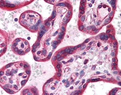
Human Placenta (formalin-fixed, paraffin-embedded) stained with HIF3A antibody (GTX85702) at 5 ug/ml followed by biotinylated goat anti-rabbit IgG secondary antibody LS-D1, alkaline phosphatase-streptavidin and chromogen.
![Anti-Hif Antibody - Western Blot. Western blot of Affinity Purified anti-Hif antibody shows detection of a band ~72 kD corresponding to mouse Hif3a (arrowhead). Approximately 10 ug of a CoCl2 treated 3T3 cell lysate (lane 1) and control 3T3 cell lysate (lane 2) were separated by 4-20% SDS-PAGE and transferred onto nitrocellulose. Treatment of exponentially growing 3T3 cells with 130 uM CoCl2 for 6 h at 37 degree C effectively mimics hypoxia. After blocking the membrane was probed overnight at 4C with the primary antibody diluted to 1:1600. The membrane was washed and reacted with a 1:10000 dilution of IRDye800 conjugated Gt-a-Rabbit IgG [H&L] MX ( for 45 min at room temperature. IRDye800 fluorescence image was captured using the Odyssey Infrared Imaging System developed by LI-COR. IRDye is a trademark of LI-COR, Inc. Other detection systems will yield similar results.](https://www.genetex.com/upload/website/prouct_img/normal/GTX85702/HIF3A-antibody_WB_GTX85702-2_w_23061502_912.webp "Anti-Hif Antibody - Western Blot. Western blot of Affinity Purified anti-Hif antibody shows detection of a band ~72 kD corresponding to mouse Hif3a (arrowhead). Approximately 10 ug of a CoCl2 treated 3T3 cell lysate (lane 1) and control 3T3 cell lysate (lane 2) were separated by 4-20% SDS-PAGE and transferred onto nitrocellulose. Treatment of exponentially growing 3T3 cells with 130 uM CoCl2 for 6 h at 37 degree C effectively mimics hypoxia. After blocking the membrane was probed overnight at 4C with the primary antibody diluted to 1:1600. The membrane was washed and reacted with a 1:10000 dilution of IRDye800 conjugated Gt-a-Rabbit IgG [H&L] MX ( for 45 min at room temperature. IRDye800 fluorescence image was captured using the Odyssey Infrared Imaging System developed by LI-COR. IRDye is a trademark of LI-COR, Inc. Other detection systems will yield similar results.")
 stained with HIF3A antibody (GTX85702) at 5 ug/ml followed by biotinylated goat anti-rabbit IgG secondary antibody LS-D1, alkaline phosphatase-streptavidin and chromogen.")
 and untreated (lane 2) NIH-3T3 whole cell lysate using GTX85702 HIF3 alpha antibody. Loading : 10 μg Dilution : 1:1600")
Human Placenta (formalin-fixed, paraffin-embedded) stained with HIF3A antibody (GTX85702) at 5 ug/ml followed by biotinylated goat anti-rabbit IgG secondary antibody LS-D1, alkaline phosphatase-streptavidin and chromogen.
HIF3 alpha antibody
GTX85702
ApplicationsWestern Blot, ELISA, ImmunoHistoChemistry, ImmunoHistoChemistry Paraffin
Product group Antibodies
ReactivityHuman, Mouse
TargetHif3a
Overview
- SupplierGeneTex
- Product NameHIF3 alpha antibody
- Delivery Days Customer9
- Application Supplier NoteWB: 1:1000-1:8000. ELISA: 1:14000-1:80000. *Optimal dilutions/concentrations should be determined by the researcher.Not tested in other applications.
- ApplicationsWestern Blot, ELISA, ImmunoHistoChemistry, ImmunoHistoChemistry Paraffin
- CertificationResearch Use Only
- ClonalityPolyclonal
- Concentration1.2 mg/ml
- ConjugateUnconjugated
- Gene ID53417
- Target nameHif3a
- Target descriptionhypoxia inducible factor 3, alpha subunit
- Target synonymsIpas, MOP7, NEPAS, bHLHe17, hypoxia-inducible factor 3-alpha, HIF-3-alpha, HIF3-alpha, HIF3-alpha-1, basic-helix-loop-helix-PAS protein MOP7, hypoxia inducible factor three alpha, inhibitory PAS domain protein, member of PAS protein 7, neonatal and embryonic PAS protein
- HostRabbit
- IsotypeIgG
- Protein IDQ0VBL6
- Protein NameHypoxia-inducible factor 3-alpha
- ReactivityHuman, Mouse
- Storage Instruction-20°C or -80°C,2°C to 8°C
- UNSPSC12352203
References
- Qu X, Li H, Meng L. XBP1 Regulates the Transcription of HIF-1a in BALB/c Mice with Chronic Rhinosinusitis without Polyps. Anal Cell Pathol (Amst). 2022,2022:3066456. doi: 10.1155/2022/3066456Read this paper
- Sun DG, Tian S, Zhang L, et al. The miRNA-29b Is Downregulated in Placenta During Gestational Diabetes Mellitus and May Alter Placenta Development by Regulating Trophoblast Migration and Invasion Through a HIF3A-Dependent Mechanism. Front Endocrinol (Lausanne). 2020,11:169. doi: 10.3389/fendo.2020.00169Read this paper
Datasheet
Related products
Product group Antibodies

Goat anti-HIF3A AntibodyEB10073
ApplicationsELISA, ImmunoHistoChemistry
ReactivityHuman, Mouse, Rat
TargetHif3a
- SizePrice