

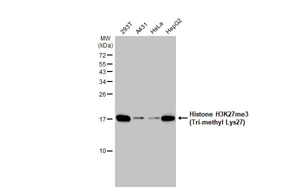
Various whole cell extracts (30 μg) were separated by 15% SDS-PAGE, and the membrane was blotted with Histone H3K27me3 (Tri-methyl Lys27) antibody (GTX121184) diluted at 1:1000. The HRP-conjugated anti-rabbit IgG antibody (GTX213110-01) was used to detect the primary antibody, and the signal was developed with Trident ECL plus-Enhanced.
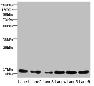
 were separated by 15% SDS-PAGE, and the membrane was blotted with Histone H3K27me3 (Tri-methyl Lys27) antibody (GTX121184) diluted at 1:1000. The HRP-conjugated anti-rabbit IgG antibody (GTX213110-01) was used to detect the primary antibody, and the signal was developed with Trident ECL plus-Enhanced.")
 were separated by 15% SDS-PAGE, and the membrane was blotted with Histone H3K27me3 (Tri-methyl Lys27) antibody (GTX121184) diluted at 1:1000. The HRP-conjugated anti-rabbit IgG antibody (GTX213110-01) was used to detect the primary antibody.")
 antibody (GTX121184) diluted at 1:200. The signal was developed using goat anti-rabbit IgG antibody (Dylight594) (GTX213110-05). Blue: Nuclear staining with Hoechst 33342.
Antigen Retrieval: Trilogy? (EDTA based, pH 8.0) buffer, 15min")
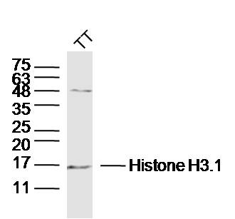
 antibody (GTX121184) diluted at 1:2500.")
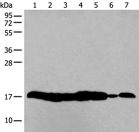
 were separated by 15% SDS-PAGE, and the membrane was blotted with Histone H3K27me3 (trimethyl Lys27) antibody (GTX121184) diluted at 1:1000.")
![Histone H3K27me3 (Tri-methyl Lys27) antibody detects Histone H3K27me3 (Tri-methyl Lys27) protein at nucleus by immunofluorescent analysis. Sample: 293T cells were fixed in 4% paraformaldehyde at RT for 15 min. Green: Histone H3K27me3 (Tri-methyl Lys27) stained by Histone H3K27me3 (Tri-methyl Lys27) antibody (GTX121184) diluted at 1:500. Red: alpha Tubulin, a cytoskeleton marker, stained by alpha Tubulin antibody [GT114] (GTX628802) diluted at 1:1000.](https://www.genetex.com/upload/website/prouct_img/normal/GTX121184/GTX121184_44552_20220429_ICC_IF_w_23060519_989.webp "Histone H3K27me3 (Tri-methyl Lys27) antibody detects Histone H3K27me3 (Tri-methyl Lys27) protein at nucleus by immunofluorescent analysis. Sample: 293T cells were fixed in 4% paraformaldehyde at RT for 15 min. Green: Histone H3K27me3 (Tri-methyl Lys27) stained by Histone H3K27me3 (Tri-methyl Lys27) antibody (GTX121184) diluted at 1:500. Red: alpha Tubulin, a cytoskeleton marker, stained by alpha Tubulin antibody [GT114] (GTX628802) diluted at 1:1000.")
 antibody detects Histone H3K27me3 (trimethyl Lys27) protein on zebrafish by whole mount immunohistochemical analysis. Sample: Paraformaldehyde-fixed 2 day-post-fertilization zebrafish embryo. Histone H3K27me3 (trimethyl Lys27) antibody (GTX121184) dilution: 1:100.")
(GTX121184) antibody at 1:500 dilution.
Antigen Retrieval: Trilogy? (EDTA based, pH 8.0) buffer, 15min")
![Histone H3K27me3 (Tri-methyl Lys27) antibody detects Histone H3K27me3 (Tri-methyl Lys27) protein at nucleus by immunofluorescent analysis. Sample: 293T cells were fixed in 4% paraformaldehyde at RT for 15 min. Green: Histone H3K27me3 (Tri-methyl Lys27) stained by Histone H3K27me3 (Tri-methyl Lys27) antibody (GTX121184) diluted at 1:500. Red: alpha Tubulin, a cytoskeleton marker, stained by alpha Tubulin antibody [GT114] (GTX628802) diluted at 1:1000.](https://www.genetex.com/upload/website/prouct_img/normal/GTX121184/GTX121184_44510_20220318_ICC_IF_w_23060519_770.webp "Histone H3K27me3 (Tri-methyl Lys27) antibody detects Histone H3K27me3 (Tri-methyl Lys27) protein at nucleus by immunofluorescent analysis. Sample: 293T cells were fixed in 4% paraformaldehyde at RT for 15 min. Green: Histone H3K27me3 (Tri-methyl Lys27) stained by Histone H3K27me3 (Tri-methyl Lys27) antibody (GTX121184) diluted at 1:500. Red: alpha Tubulin, a cytoskeleton marker, stained by alpha Tubulin antibody [GT114] (GTX628802) diluted at 1:1000.")
Various whole cell extracts (30 μg) were separated by 15% SDS-PAGE, and the membrane was blotted with Histone H3K27me3 (Tri-methyl Lys27) antibody (GTX121184) diluted at 1:1000. The HRP-conjugated anti-rabbit IgG antibody (GTX213110-01) was used to detect the primary antibody, and the signal was developed with Trident ECL plus-Enhanced.
Histone H3K27me3 (Tri-methyl Lys27) antibody
GTX121184
ApplicationsDot Blot, Electron Microscopy, ImmunoFluorescence, Western Blot, ImmunoCytoChemistry, ImmunoHistoChemistry, ImmunoHistoChemistry Frozen, ImmunoHistoChemistry Paraffin
Product group Antibodies
ReactivityHuman, Mouse, Rat, Zebra Fish
Overview
- SupplierGeneTex
- Product NameHistone H3K27me3 (Tri-methyl Lys27) antibody
- Delivery Days Customer9
- Application Supplier NoteWB: 1:500-1:3000. ICC/IF: 1:100-1:1000. IHC-P: 1:100-1:1000. IHC-Fr: 1:100-1:1000. IHC-Wm: 1:100-1:500. *Optimal dilutions/concentrations should be determined by the researcher.Not tested in other applications.
- ApplicationsDot Blot, Electron Microscopy, ImmunoFluorescence, Western Blot, ImmunoCytoChemistry, ImmunoHistoChemistry, ImmunoHistoChemistry Frozen, ImmunoHistoChemistry Paraffin
- CertificationResearch Use Only
- ClonalityPolyclonal
- Concentration1 mg/ml
- ConjugateUnconjugated
- HostRabbit
- IsotypeIgG
- ReactivityHuman, Mouse, Rat, Zebra Fish
- Storage Instruction-20°C or -80°C,2°C to 8°C
- UNSPSC41116161
Datasheet
Related products
Product group Antibodies
Anti-HIST1H3A AntibodyA44415
ApplicationsWestern Blot, ImmunoHistoChemistry
ReactivityHuman, Mouse
- SizePrice

Product group Antibodies
Anti-Histone H3 HIST1H3A/B/C/D/E/F/G/H/I/J Antibody Picoband(r)A12477-2-CARRIER-FREE
ApplicationsFlow Cytometry, ImmunoFluorescence, Western Blot, ELISA, ImmunoCytoChemistry, ImmunoHistoChemistry
ReactivityHuman, Mouse, Rat
TargetH3C1
- SizePrice
Product group Antibodies
ApplicationsImmunoFluorescence, ImmunoPrecipitation, Western Blot, ChIP Chromatin ImmunoPrecipitation, ImmunoHistoChemistry
ReactivityHuman, Mouse, Rat, Other Species
TargetH3C1
- SizePrice

Product group Antibodies
Anti-HIST1H3A AntibodyAMAB91331
ApplicationsWestern Blot, ImmunoCytoChemistry, ImmunoHistoChemistry
ReactivityHuman, Mouse, Rat
TargetH3C1
- SizePrice

Product group Antibodies

HIST1H3A AntibodyLS-C763756
ApplicationsImmunoFluorescence, Western Blot, ELISA, ImmunoHistoChemistry, ImmunoHistoChemistry Paraffin
ReactivityHuman, Mouse, Rat
TargetH3C1
- SizePrice
Product group Antibodies
References
Histone H3.1 Polyclonal AntibodyBS-17422R
ApplicationsFlow Cytometry, ImmunoFluorescence, Western Blot, ELISA, ImmunoCytoChemistry, ImmunoHistoChemistry, ImmunoHistoChemistry Frozen, ImmunoHistoChemistry Paraffin
ReactivityBovine, Equine, Human, Mouse, Porcine, Primate, Rabbit, Rat, Sheep
TargetH3C1
- SizePrice
Product group Antibodies
HIST1H3A AntibodyCSB-PA010418ESR1HU
ApplicationsWestern Blot, ELISA, ImmunoHistoChemistry
ReactivityHuman, Mouse
TargetH3C1
- SizePrice
Product group Antibodies
ApplicationsELISA, ImmunoCytoChemistry
TargetH3C1
- SizePrice