

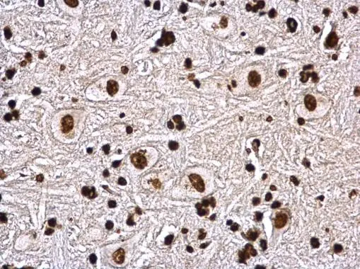
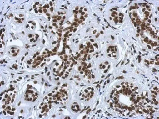
Histone H4 antibody detects Histone H4 protein at nucleus on rat brain stem by immunohistochemical analysis. Sample: Paraffin-embedded rat brain stem. Histone H4 antibody (GTX129560) dilution: 1:500.
Antigen Retrieval: Trilogy? (EDTA based, pH 8.0) buffer, 15min
 dilution: 1:500.
Antigen Retrieval: Trilogy? (EDTA based, pH 8.0) buffer, 15min")
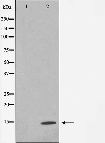
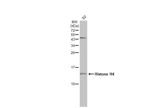
 were separated by 15% SDS-PAGE, and the membrane was blotted with Histone H4 antibody (GTX129560) diluted at a dilution of 1:5000.")
 were separated by 15% SDS-PAGE, and the membrane was blotted with Histone H4 antibody (GTX129560) diluted at 1:1000.")
 dilution: 1:500.
Antigen Retrieval: Trilogy? (EDTA based, pH 8.0) buffer, 15min")
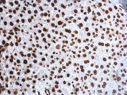
![Histone H4 antibody detects Histone H4 protein at nucleus by immunofluorescent analysis. Sample: HeLa cells were fixed in 4% paraformaldehyde at RT for 15 min. Green: Histone H4 protein stained by Histone H4 antibody (GTX129560) diluted at 1:5000. Red: alpha Tubulin, a cytoskeleton marker, stained by alpha Tubulin antibody [GT114] (GTX628802) diluted at 1:2000. Blue: Hoechst 33342 staining.](https://www.genetex.com/upload/website/prouct_img/normal/GTX129560/GTX129560_41423_IFA_w_23060523_532.webp "Histone H4 antibody detects Histone H4 protein at nucleus by immunofluorescent analysis. Sample: HeLa cells were fixed in 4% paraformaldehyde at RT for 15 min. Green: Histone H4 protein stained by Histone H4 antibody (GTX129560) diluted at 1:5000. Red: alpha Tubulin, a cytoskeleton marker, stained by alpha Tubulin antibody [GT114] (GTX628802) diluted at 1:2000. Blue: Hoechst 33342 staining.")
 dilution: 1:500.
Antigen Retrieval: Trilogy? (EDTA based, pH 8.0) buffer, 15min")
Histone H4 antibody detects Histone H4 protein at nucleus on rat brain stem by immunohistochemical analysis. Sample: Paraffin-embedded rat brain stem. Histone H4 antibody (GTX129560) dilution: 1:500.
Antigen Retrieval: Trilogy? (EDTA based, pH 8.0) buffer, 15min
Histone H4 antibody
GTX129560
ApplicationsImmunoFluorescence, Western Blot, ImmunoCytoChemistry, ImmunoHistoChemistry, ImmunoHistoChemistry Paraffin
Product group Antibodies
ReactivityHuman, Mouse, Rat, Zebra Fish
TargetH4C1
Overview
- SupplierGeneTex
- Product NameHistone H4 antibody
- Delivery Days Customer9
- Application Supplier NoteWB: 1:500-1:10000. ICC/IF: 1:100-1:5000. IHC-P: 1:100-1:1000. *Optimal dilutions/concentrations should be determined by the researcher.Not tested in other applications.
- ApplicationsImmunoFluorescence, Western Blot, ImmunoCytoChemistry, ImmunoHistoChemistry, ImmunoHistoChemistry Paraffin
- CertificationResearch Use Only
- ClonalityPolyclonal
- Concentration1.56 mg/ml
- ConjugateUnconjugated
- Gene ID8359
- Target nameH4C1
- Target descriptionH4 clustered histone 1
- Target synonymsH4-16, H4C11, H4C12, H4C13, H4C14, H4C15, H4C16, H4C2, H4C3, H4C4, H4C5, H4C6, H4C8, H4C9, H4FA, HIST1H4A, histone H4, H4 histone family, member A, histone 1, H4a, histone cluster 1 H4 family member a, histone cluster 1, H4a
- HostRabbit
- IsotypeIgG
- ReactivityHuman, Mouse, Rat, Zebra Fish
- Storage Instruction-20°C or -80°C,2°C to 8°C
- UNSPSC12352203
References
- Guo Y, Chomiak AA, Hong Y, et al. Histone H2A ubiquitination resulting from Brap loss of function connects multiple aging hallmarks and accelerates neurodegeneration. iScience. 2022,25(7):104519. doi: 10.1016/j.isci.2022.104519Read this paper
- Chomiak AA, Guo Y, Kopsidas CA, et al. Nde1 is required for heterochromatin compaction and stability in neocortical neurons. iScience. 2022,25(6):104354. doi: 10.1016/j.isci.2022.104354Read this paper
- Liu MK, Lin JJ, Chen CY, et al. Topoisomerase II Inhibitors Can Enhance Baculovirus-Mediated Gene Expression in Mammalian Cells through the DNA Damage Response. Int J Mol Sci. 2016,17(6). doi: 10.3390/ijms17060931Read this paper
Datasheet
Related products
Product group Antibodies
ApplicationsImmunoPrecipitation, Western Blot, ImmunoHistoChemistry
ReactivityHuman, Mouse, Rat
- SizePrice
Product group Antibodies
ApplicationsImmunoFluorescence, Western Blot, ImmunoCytoChemistry, ImmunoHistoChemistry, ImmunoHistoChemistry Paraffin
ReactivityHuman, Mouse, Rat
TargetH4C1
- SizePrice
Product group Antibodies
References
ApplicationsImmunoFluorescence, Western Blot, ImmunoCytoChemistry, ImmunoHistoChemistry, ImmunoHistoChemistry Paraffin
ReactivityHuman, Mouse, Rat
TargetH4C1
- SizePrice
Product group Antibodies
ApplicationsDot Blot, ImmunoFluorescence, ImmunoPrecipitation, Western Blot, ChIP Chromatin ImmunoPrecipitation, ImmunoCytoChemistry, ImmunoHistoChemistry, ImmunoHistoChemistry Paraffin
ReactivityHuman, Mouse, Rat
TargetH4C1
- SizePrice
Product group Antibodies
Histone H4 antibodyGTX52365
ApplicationsImmunoFluorescence, Western Blot, ImmunoCytoChemistry, ImmunoHistoChemistry
ReactivityHuman, Monkey, Mouse, Rat
TargetH4C1
- SizePrice
Product group Antibodies

HIST1H4A Antibody (aa1-103)LS-C375024
ApplicationsWestern Blot
ReactivityBovine, Human, Mouse, Rat
TargetH4C1
- SizePrice
Product group Antibodies
References
ApplicationsDot Blot, ImmunoFluorescence, Western Blot, ImmunoCytoChemistry
ReactivityHuman, Mouse
TargetH4C1
- SizePrice
Product group Antibodies
ApplicationsDot Blot, ImmunoFluorescence, Western Blot, ImmunoCytoChemistry, ImmunoHistoChemistry, ImmunoHistoChemistry Paraffin
ReactivityHuman, Mouse, Rat
TargetH4C1
- SizePrice
Product group Antibodies
Histone H4 antibodyGTX129561
ApplicationsImmunoFluorescence, Western Blot, ImmunoCytoChemistry, ImmunoHistoChemistry, ImmunoHistoChemistry Paraffin
ReactivityDrosophila, Human, Mouse
TargetH4C1
- SizePrice
Product group Antibodies
References
ApplicationsDot Blot, ImmunoFluorescence, Western Blot, ImmunoCytoChemistry, ImmunoHistoChemistry, ImmunoHistoChemistry Paraffin
ReactivityHuman, Mouse, Rat
TargetH4C1
- SizePrice