

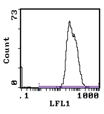
Staining of peripheral blood lymphocytes using HLA Class I antibody (GTX27855).
Staining of peripheral blood lymphocytes using HLA Class I antibody (GTX27855).
HLA Class I antibody [W6/32]
GTX27855
ApplicationsFlow Cytometry, ImmunoFluorescence, ELISA, ImmunoCytoChemistry, ImmunoHistoChemistry, ImmunoHistoChemistry Frozen
Product group Antibodies
ReactivityHuman
Overview
- SupplierGeneTex
- Product NameHLA Class I antibody [W6/32]
- Delivery Days Customer9
- Application Supplier NoteFor IHC(frozen) a three-step avidin-biotin complex system, a dilution of 1:50 to 1:100 may be used as a guideline. In general, the 0.05M Borate pH 8.0 containing 0.15M Sodium Chloride, 0.05% Sodium Azide, is a good dilutent to use with most antibodies. Optimal dilutions/concentrations should be determined by the researcher.
- ApplicationsFlow Cytometry, ImmunoFluorescence, ELISA, ImmunoCytoChemistry, ImmunoHistoChemistry, ImmunoHistoChemistry Frozen
- CertificationResearch Use Only
- ClonalityMonoclonal
- Clone IDW6/32
- ConjugateUnconjugated
- HostMouse
- IsotypeIgG2a
- Scientific DescriptionHLA-class I antigens are widely distributed on human nucleated cells. The intensity of HLA-ABC maybe altered in pathological states e.g. malignant cells may loose HLA-ABC, whereas hepatocytes in alcoholic hepatitis, biliary cirrhosis and chronic active hepatitis may show enhanced expression of HLA-ABC.
- ReactivityHuman
- Storage Instruction2°C to 8°C
- UNSPSC41116161
Datasheet
Related products
Product group Antibodies
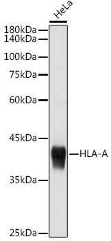
HLA-A AntibodyCSB-PA398166
ApplicationsWestern Blot, ELISA
ReactivityHuman
TargetHLA-A
- SizePrice
Product group Antibodies

Anti-HLA-class I [YTH 862.2]Ab00200-1.1
ApplicationsFlow Cytometry, ImmunoFluorescence, ImmunoPrecipitation
ReactivityHuman
TargetHLA-A
- SizePrice

Product group Antibodies

HLA-A AntibodyLS-C482358
ApplicationsImmunoFluorescence, Western Blot, ImmunoCytoChemistry, ImmunoHistoChemistry, ImmunoHistoChemistry Paraffin
ReactivityHuman, Mouse, Rat
TargetHLA-A
- SizePrice
Product group Antibodies

Mouse anti HLA Class IMUB2035P
ApplicationsFlow Cytometry, ImmunoHistoChemistry, ImmunoHistoChemistry Frozen
ReactivityHuman
- SizePrice
Product group Antibodies
HLA Class I antibody [MEM-81]GTX23975
ApplicationsFlow Cytometry
ReactivityHuman
TargetHLA-A
- SizePrice
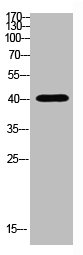
![WB analysis of Ramos cell lysate (1% laurylmaltoside - non-reduced sample) using GTX29090 HLA Class I antibody [MEM-147].](https://www.genetex.com/upload/website/prouct_img/normal/GTX29090/GTX29090_20191028_WB_1_w_23060722_830.webp)
Product group Antibodies
HLA Class I antibody [MEM-147]GTX29090
ApplicationsFlow Cytometry, ImmunoPrecipitation, Western Blot
ReactivityHuman
TargetHLA-A
- SizePrice
Product group Antibodies
HLA-A antibodyGTX54099
ApplicationsWestern Blot, ImmunoHistoChemistry, ImmunoHistoChemistry Paraffin
ReactivityHuman, Mouse
TargetHLA-A
- SizePrice