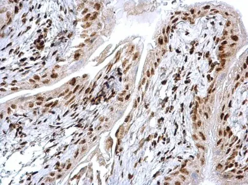
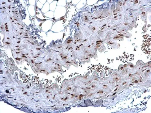
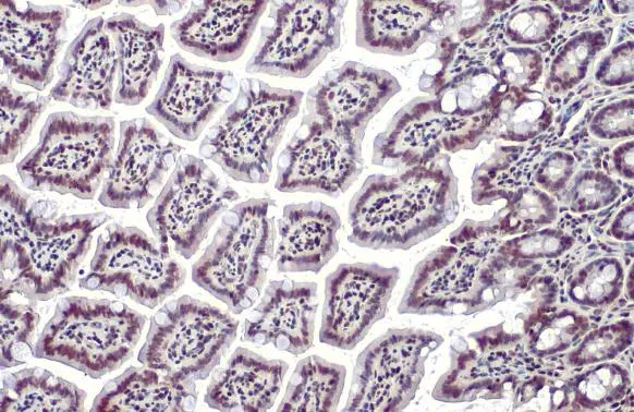
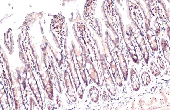
HMGB1 antibody detects HMGB1 protein at nucleus on mouse vein by immunohistochemical analysis. Sample: Paraffin-embedded mouse vein. HMGB1 antibody (GTX127344) dilution: 1:500.
Antigen Retrieval: Trilogy? (EDTA based, pH 8.0) buffer, 15min
HMGB1 antibody
GTX127344
ApplicationsImmunoFluorescence, ImmunoPrecipitation, Western Blot, ImmunoCytoChemistry, ImmunoHistoChemistry, ImmunoHistoChemistry Frozen, ImmunoHistoChemistry Paraffin, Other Application
Product group Antibodies
ReactivityHuman, Mouse, Rat
TargetHMGB1
Overview
- SupplierGeneTex
- Product NameHMGB1 antibody
- Delivery Days Customer9
- Application Supplier NoteWB: 1:500-1:3000. ICC/IF: 1:100-1:1000. IHC-P: 1:100-1:1000. IHC-Fr: 1:100-1:1000. IP: 1:100-1:500. *Optimal dilutions/concentrations should be determined by the researcher.Not tested in other applications.
- ApplicationsImmunoFluorescence, ImmunoPrecipitation, Western Blot, ImmunoCytoChemistry, ImmunoHistoChemistry, ImmunoHistoChemistry Frozen, ImmunoHistoChemistry Paraffin, Other Application
- CertificationResearch Use Only
- ClonalityPolyclonal
- Concentration0.17 mg/ml
- ConjugateUnconjugated
- Gene ID3146
- Target nameHMGB1
- Target descriptionhigh mobility group box 1
- Target synonymsHMG-1, HMG1, HMG3, SBP-1, high mobility group protein B1, Amphoterin, Sulfoglucuronyl carbohydrate binding protein, high-mobility group (nonhistone chromosomal) protein 1
- HostRabbit
- IsotypeIgG
- Protein IDP09429
- Protein NameHigh mobility group protein B1
- Scientific DescriptionDNA binding proteins that associates with chromatin and has the ability to bend DNA. Binds preferentially single-stranded DNA. Involved in V(D)J recombination by acting as a cofactor of the RAG complex. Acts by stimulating cleavage and RAG protein binding at the 23 bp spacer of conserved recombination signal sequences (RSS). Heparin-binding protein that has a role in the extension of neurite-type cytoplasmic processes in developing cells.
- ReactivityHuman, Mouse, Rat
- Storage Instruction-20°C or -80°C,2°C to 8°C
- UNSPSC12352203
References
- Li L, Li F, Bai X, et al. Circulating extracellular vesicles from patients with traumatic brain injury induce cerebrovascular endothelial dysfunction. Pharmacol Res. 2023,192:106791. doi: 10.1016/j.phrs.2023.106791Read this paper
- Kubota R, Hayashi N, Kinoshita K, et al. Inhibition of γ-glutamyltransferase ameliorates ischaemia-reoxygenation tissue damage in rats with hepatic steatosis. Br J Pharmacol. 2020,177(22):5195-5207. doi: 10.1111/bph.15258Read this paper


![HMGB1 antibody detects HMGB1 Protein expression by immunohistochemical analysis. Sample: Frozen-sectioned adult mouse cerebellum. Green: HMGB1 stained by HMGB1 antibody (GTX127344) diluted at 1:250. Red: NF-H, stained by NF-H antibody [GT114] (GTX634289) diluted at 1:500. Blue: Fluoroshield with DAPI (GTX30920).
Antigen Retrieval: Citrate buffer, pH 6.0, 5 min HMGB1 antibody detects HMGB1 Protein expression by immunohistochemical analysis. Sample: Frozen-sectioned adult mouse cerebellum. Green: HMGB1 stained by HMGB1 antibody (GTX127344) diluted at 1:250. Red: NF-H, stained by NF-H antibody [GT114] (GTX634289) diluted at 1:500. Blue: Fluoroshield with DAPI (GTX30920).
Antigen Retrieval: Citrate buffer, pH 6.0, 5 min](https://www.genetex.com/upload/website/prouct_img/normal/GTX127344/GTX127344_40905_20170829_IHC-Fr_M_w_23060522_266.webp)
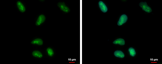
![HMGB1 antibody detects HMGB1 protein at nucleus by immunofluorescent analysis. Sample: DIV9 rat E18 primary cortical neurons were fixed in 4% paraformaldehyde at RT for 15 min. Green: HMGB1 protein stained by HMGB1 antibody (GTX127344) diluted at 1:500. Red: beta Tubulin 3/ Tuj1, a neuron cell marker, stained by beta Tubulin 3/ Tuj1 antibody [GT11710] (GTX631836) diluted at 1:500. HMGB1 antibody detects HMGB1 protein at nucleus by immunofluorescent analysis. Sample: DIV9 rat E18 primary cortical neurons were fixed in 4% paraformaldehyde at RT for 15 min. Green: HMGB1 protein stained by HMGB1 antibody (GTX127344) diluted at 1:500. Red: beta Tubulin 3/ Tuj1, a neuron cell marker, stained by beta Tubulin 3/ Tuj1 antibody [GT11710] (GTX631836) diluted at 1:500.](https://www.genetex.com/upload/website/prouct_img/normal/GTX127344/GTX127344_40905_20170727_IFA_R_w_23060522_760.webp)
![HMGB1 antibody detects HMGB1 protein at nucleus by immunofluorescent analysis. Sample: HeLa cells were fixed in 4% paraformaldehyde at RT for 15 min. Green: HMGB1 stained by HMGB1 antibody (GTX127344) diluted at 1:500. Red: alpha Tubulin, a cytoskeleton marker, stained by alpha Tubulin antibody [GT114] (GTX628802) diluted at 1:1000. HMGB1 antibody detects HMGB1 protein at nucleus by immunofluorescent analysis. Sample: HeLa cells were fixed in 4% paraformaldehyde at RT for 15 min. Green: HMGB1 stained by HMGB1 antibody (GTX127344) diluted at 1:500. Red: alpha Tubulin, a cytoskeleton marker, stained by alpha Tubulin antibody [GT114] (GTX628802) diluted at 1:1000.](https://www.genetex.com/upload/website/prouct_img/normal/GTX127344/GTX127344_44307_20220121_ICC_IF_w_23060522_466.webp)

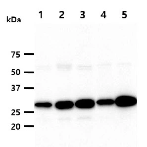
![HMGB1 antibody [GT383] detects HMGB1 protein by western blot analysis. A. 30 μg 293T whole cell lysate/extract B. 30 μg A431 whole cell lysate/extract C. 30 μg HeLa whole cell lysate/extract D. 30 μg HepG2 whole cell lysate/extract E. 30 μg A375 whole cell lysate/extract 12% SDS-PAGE HMGB1 antibody [GT383] (GTX628834) dilution: 1:1000 The HRP-conjugated anti-mouse IgG antibody (GTX213111-01) was used to detect the primary antibody.](https://www.genetex.com/upload/website/prouct_img/normal/GTX628834/GTX628834_41225_WB_w_23061202_196.webp)
![HMGB1 antibody [GT348] detects HMGB1 protein by western blot analysis. A. 30 μg 293T whole cell lysate/extract B. 30 μg A431 whole cell lysate/extract C. 30 μg HepG2 whole cell lysate/extract 12% SDS-PAGE HMGB1 antibody [GT348] (GTX628835) dilution: 1:1000 The HRP-conjugated anti-mouse IgG antibody (GTX213111-01) was used to detect the primary antibody.](https://www.genetex.com/upload/website/prouct_img/normal/GTX628835/GTX628835_41225_WB_w_23061202_883.webp)
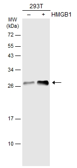
![Non-transfected (–) and transfected (+) 293T whole cell extracts (30 μg) were separated by 12% SDS-PAGE, and the membrane was blotted with HMGB1 antibody [GT412] (GTX629400) diluted at 1:3000. The HRP-conjugated anti-mouset IgG antibody (GTX213111-01) was used to detect the primary antibody.](https://www.genetex.com/upload/website/prouct_img/normal/GTX629400/GTX629400_41323_20181005_WB_B_w_23061202_582.webp)
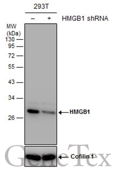
![Non-transfected (–) and transfected (+) 293T whole cell extracts (30 μg) were separated by 12% SDS-PAGE, and the membrane was blotted with HMGB1 antibody [GT349] (GTX629403) diluted at 1:5000.](https://www.genetex.com/upload/website/prouct_img/normal/GTX629403/GTX629403_41323_20160602_WB_shRNA_watermark_w_23061202_915.webp)