

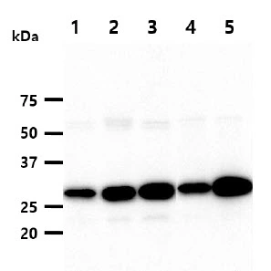
WB analysis of various samples using GTX57549 HMGB1 antibody. Lane 1 : HeLa whole cell lysate Lane 2 : Jurkat whole cell lysate Lane 3 : K562 whole cell lysate Lane 4 : A549 whole cell lysate Lane 5 : MCF-7 whole cell lysate Loading : 40 μg Dilution : 1:1000

WB analysis of various samples using GTX57549 HMGB1 antibody. Lane 1 : HeLa whole cell lysate Lane 2 : Jurkat whole cell lysate Lane 3 : K562 whole cell lysate Lane 4 : A549 whole cell lysate Lane 5 : MCF-7 whole cell lysate Loading : 40 μg Dilution : 1:1000
HMGB1 antibody [J2E1]
GTX57549
ApplicationsWestern Blot, ImmunoHistoChemistry, ImmunoHistoChemistry Paraffin
Product group Antibodies
ReactivityHuman
TargetHMGB1
Overview
- SupplierGeneTex
- Product NameHMGB1 antibody [J2E1]
- Delivery Days Customer9
- Application Supplier NoteWB: 1:500-1:2000. IHC-P: 1:100-1:300. *Optimal dilutions/concentrations should be determined by the researcher.Not tested in other applications.
- ApplicationsWestern Blot, ImmunoHistoChemistry, ImmunoHistoChemistry Paraffin
- CertificationResearch Use Only
- ClonalityMonoclonal
- Clone IDJ2E1
- Concentration1 mg/ml
- ConjugateUnconjugated
- Gene ID3146
- Target nameHMGB1
- Target descriptionhigh mobility group box 1
- Target synonymsHMG-1, HMG1, HMG3, SBP-1, high mobility group protein B1, Amphoterin, Sulfoglucuronyl carbohydrate binding protein, high-mobility group (nonhistone chromosomal) protein 1
- HostMouse
- IsotypeIgG2b
- Protein IDP09429
- Protein NameHigh mobility group protein B1
- Scientific DescriptionThis gene encodes a protein that belongs to the High Mobility Group-box superfamily. The encoded non-histone, nuclear DNA-binding protein regulates transcription, and is involved in organization of DNA. This protein plays a role in several cellular processes, including inflammation, cell differentiation and tumor cell migration. Multiple pseudogenes of this gene have been identified. Alternative splicing results in multiple transcript variants that encode the same protein. [provided by RefSeq, Sep 2015]
- ReactivityHuman
- Storage Instruction-20°C or -80°C,2°C to 8°C
- UNSPSC12352203
Datasheet
Related products
Product group Antibodies
Anti-HMGB1 Antibody144-02553
ApplicationsImmunoFluorescence, Western Blot, ImmunoHistoChemistry
ReactivityHuman, Mouse, Rat
TargetHMGB1
- SizePrice

Product group Antibodies
Anti-HMGB1 Antibody Picoband(r)A00066-1-CARRIER-FREE
ApplicationsFlow Cytometry, ImmunoFluorescence, Western Blot, ImmunoCytoChemistry, ImmunoHistoChemistry
ReactivityHuman, Mouse, Rat
TargetHMGB1
- SizePrice
Product group Antibodies
References
HMGB1 antibodyGTX127344
ApplicationsImmunoFluorescence, ImmunoPrecipitation, Western Blot, ImmunoCytoChemistry, ImmunoHistoChemistry, ImmunoHistoChemistry Frozen, ImmunoHistoChemistry Paraffin, Other Application
ReactivityHuman, Mouse, Rat
TargetHMGB1
- SizePrice
Product group Antibodies
References
HMGB1 antibodyGTX101277
ApplicationsImmunoFluorescence, Western Blot, ImmunoCytoChemistry, ImmunoHistoChemistry, ImmunoHistoChemistry Paraffin
ReactivityHuman, Mouse, Porcine, Rat, Zebra Fish
TargetHMGB1
- SizePrice
Product group Antibodies
References
HMGB1 antibodyGTX112959
ApplicationsImmunoFluorescence, Western Blot, ImmunoCytoChemistry, ImmunoHistoChemistry, ImmunoHistoChemistry Paraffin
ReactivityHuman, Mouse, Rat, Zebra Fish
TargetHMGB1
- SizePrice
![HMGB1 antibody [GT383] detects HMGB1 protein by western blot analysis. A. 30 μg 293T whole cell lysate/extract B. 30 μg A431 whole cell lysate/extract C. 30 μg HeLa whole cell lysate/extract D. 30 μg HepG2 whole cell lysate/extract E. 30 μg A375 whole cell lysate/extract 12% SDS-PAGE HMGB1 antibody [GT383] (GTX628834) dilution: 1:1000 The HRP-conjugated anti-mouse IgG antibody (GTX213111-01) was used to detect the primary antibody.](https://www.genetex.com/upload/website/prouct_img/normal/GTX628834/GTX628834_41225_WB_w_23061202_196.webp)
Product group Antibodies
References
HMGB1 antibody [GT383]GTX628834
ApplicationsImmunoFluorescence, Western Blot, ImmunoCytoChemistry, ImmunoHistoChemistry, ImmunoHistoChemistry Frozen, ImmunoHistoChemistry Paraffin
ReactivityHuman, Mouse, Rat
TargetHMGB1
- SizePrice
![HMGB1 antibody [GT348] detects HMGB1 protein by western blot analysis. A. 30 μg 293T whole cell lysate/extract B. 30 μg A431 whole cell lysate/extract C. 30 μg HepG2 whole cell lysate/extract 12% SDS-PAGE HMGB1 antibody [GT348] (GTX628835) dilution: 1:1000 The HRP-conjugated anti-mouse IgG antibody (GTX213111-01) was used to detect the primary antibody.](https://www.genetex.com/upload/website/prouct_img/normal/GTX628835/GTX628835_41225_WB_w_23061202_883.webp)
Product group Antibodies
HMGB1 antibody [GT348]GTX628835
ApplicationsImmunoFluorescence, Western Blot, ImmunoCytoChemistry, ImmunoHistoChemistry, ImmunoHistoChemistry Frozen, ImmunoHistoChemistry Paraffin
ReactivityHuman, Mouse, Rat
TargetHMGB1
- SizePrice
![Non-transfected (–) and transfected (+) 293T whole cell extracts (30 μg) were separated by 12% SDS-PAGE, and the membrane was blotted with HMGB1 antibody [GT412] (GTX629400) diluted at 1:3000. The HRP-conjugated anti-mouset IgG antibody (GTX213111-01) was used to detect the primary antibody.](https://www.genetex.com/upload/website/prouct_img/normal/GTX629400/GTX629400_41323_20181005_WB_B_w_23061202_582.webp)
Product group Antibodies
References
HMGB1 antibody [GT412]GTX629400
ApplicationsImmunoFluorescence, Western Blot, ImmunoCytoChemistry, ImmunoHistoChemistry, ImmunoHistoChemistry Paraffin
ReactivityHuman, Mouse, Rat
TargetHMGB1
- SizePrice
![Non-transfected (–) and transfected (+) 293T whole cell extracts (30 μg) were separated by 12% SDS-PAGE, and the membrane was blotted with HMGB1 antibody [GT349] (GTX629403) diluted at 1:5000.](https://www.genetex.com/upload/website/prouct_img/normal/GTX629403/GTX629403_41323_20160602_WB_shRNA_watermark_w_23061202_915.webp)
Product group Antibodies
HMGB1 antibody [GT349]GTX629403
ApplicationsImmunoFluorescence, Western Blot, ImmunoCytoChemistry, ImmunoHistoChemistry, ImmunoHistoChemistry Paraffin
ReactivityHuman, Mouse, Rat
TargetHMGB1
- SizePrice