

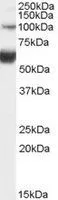
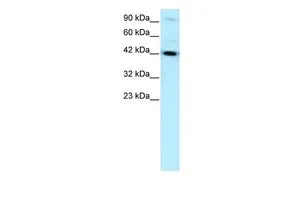
WB analysis of human liver lysate using GTX89168 HNF1 beta antibody, Internal. Dilution : 0.1μg/ml Loading : 35μg protein in RIPA buffer
WB analysis of human liver lysate using GTX89168 HNF1 beta antibody, Internal. Dilution : 0.1μg/ml Loading : 35μg protein in RIPA buffer
HNF1 beta antibody, Internal
GTX89168
ApplicationsImmunoFluorescence, Western Blot, ImmunoCytoChemistry
Product group Antibodies
ReactivityHuman
TargetHNF1B
Overview
- SupplierGeneTex
- Product NameHNF1 beta antibody, Internal
- Delivery Days Customer7
- Application Supplier NoteWB: 0.1-0.3microg/ml. *Optimal dilutions/concentrations should be determined by the researcher.Not tested in other applications.
- ApplicationsImmunoFluorescence, Western Blot, ImmunoCytoChemistry
- CertificationResearch Use Only
- ClonalityPolyclonal
- Concentration0.50 mg/ml
- ConjugateUnconjugated
- Gene ID6928
- Target nameHNF1B
- Target descriptionHNF1 homeobox B
- Target synonymsADTKD3, FJHN, HNF-1-beta, HNF-1B, HNF1beta, HNF2, HPC11, LF-B3, LFB3, MODY5, RCAD, T2D, TCF-2, TCF2, VHNF1, hepatocyte nuclear factor 1-beta, HNF1 beta A, homeoprotein LFB3, transcription factor 2, hepatic
- HostGoat
- IsotypeIgG
- Protein IDP35680
- Protein NameHepatocyte nuclear factor 1-beta
- Scientific DescriptionThis gene encodes a member of the homeodomain-containing superfamily of transcription factors. The protein binds to DNA as either a homodimer, or a heterodimer with the related protein hepatocyte nuclear factor 1-alpha. The gene has been shown to function in nephron development, and regulates development of the embryonic pancreas. Mutations in this gene result in renal cysts and diabetes syndrome and noninsulin-dependent diabetes mellitus, and expression of this gene is altered in some types of cancer. Multiple transcript variants encoding different isoforms have been found for this gene.[provided by RefSeq, Sep 2009]
- ReactivityHuman
- Storage Instruction-20°C or -80°C,2°C to 8°C
- UNSPSC12352203
Datasheet
Related products
Product group Antibodies

Anti-HNF1beta [11A1]Ab03313-1.1
ApplicationsWestern Blot, ELISA, ImmunoHistoChemistry
ReactivityHuman
TargetHNF1B
- SizePrice
Product group Antibodies
Anti-TCF2 Antibody101-11753
ApplicationsWestern Blot, ELISA
TargetHNF1B
- SizePrice
Product group Antibodies
HNF1 beta antibodyGTX129236
ApplicationsWestern Blot, ImmunoHistoChemistry, ImmunoHistoChemistry Paraffin
ReactivityHuman
TargetHNF1B
- SizePrice
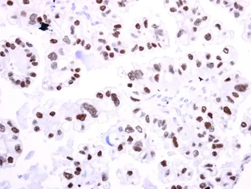
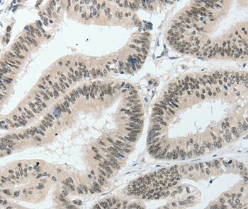
![IHC-P analysis of human liver carcinoma tissue using GTX05009 HNF1 beta antibody [HNF1B/9518]. Lower left corner : Negative control](https://www.genetex.com/upload/website/prouct_img/normal/GTX05009/GTX05009_20241212_IHC-P_24121201_143.webp)
Product group Antibodies
HNF1 beta antibody [HNF1B/9518]GTX05009
ApplicationsImmunoHistoChemistry, ImmunoHistoChemistry Paraffin
ReactivityHuman
TargetHNF1B
- SizePrice
Product group Antibodies
References
HNF1 beta antibody, N-termGTX77876
ApplicationsWestern Blot
ReactivityHuman
TargetHNF1B
- SizePrice
Product group Antibodies
ApplicationsImmunoPrecipitation, Western Blot, ImmunoCytoChemistry, ImmunoHistoChemistry
ReactivityMouse, Rat
TargetHNF1B
- SizePrice
Product group Antibodies
HNF1B Recombinant AntibodyBSM-61983R
ApplicationsWestern Blot
ReactivityHuman
TargetHNF1B
- SizePrice
Product group Antibodies
Anti-HNF1B AntibodyA46114
ApplicationsImmunoHistoChemistry
ReactivityHuman, Mouse, Rat
- SizePrice
![A549 whole cell and nuclear extracts (30 μg) were separated by 7.5% SDS-PAGE, and the membrane was blotted with HNF1 beta antibody [HL3639] (GTX641630) diluted at 1:1000. The HRP-conjugated anti-rabbit IgG antibody (GTX213110-01) was used to detect the primary antibody, and the signal was developed with Trident ECL plus-Enhanced.](https://www.genetex.com/upload/website/prouct_img/normal/GTX641630/GTX641630_T-45642_20250103_WB_Fraction_25010819_215.webp)
Product group Antibodies
HNF1 beta antibody [HL3639]GTX641630
ApplicationsWestern Blot
ReactivityHuman
TargetHNF1B
- SizePrice