

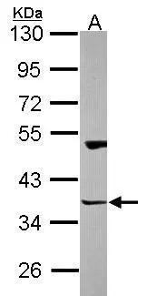
Sample (30 ug of whole cell lysate) A: NT2D1 10% SDS PAGE GTX115242 diluted at 1:1000
 antibody at 1:500 dilution.")
![Homer3 antibody detects Homer3 protein by immunofluorescent analysis. Sample: DIV9 rat E18 primary cortical neurons were fixed in 4% paraformaldehyde at RT for 15 min. Green: Homer3 protein stained by Homer3 antibody (GTX115242) diluted at 1:500. Red: beta Tubulin 3/ Tuj1, stained by beta Tubulin 3/ Tuj1 antibody [GT886] (GTX631830) diluted at 1:500. Blue: Fluoroshield with DAPI (GTX30920).](https://www.genetex.com/upload/website/prouct_img/normal/GTX115242/GTX115242_40254_20170705_IFA_R_w_23060519_624.webp "Homer3 antibody detects Homer3 protein by immunofluorescent analysis. Sample: DIV9 rat E18 primary cortical neurons were fixed in 4% paraformaldehyde at RT for 15 min. Green: Homer3 protein stained by Homer3 antibody (GTX115242) diluted at 1:500. Red: beta Tubulin 3/ Tuj1, stained by beta Tubulin 3/ Tuj1 antibody [GT886] (GTX631830) diluted at 1:500. Blue: Fluoroshield with DAPI (GTX30920).")
 dilution: 1:250.
Antigen Retrieval: Trilogy? (EDTA based, pH 8.0) buffer, 15min")

Sample (30 ug of whole cell lysate) A: NT2D1 10% SDS PAGE GTX115242 diluted at 1:1000
Homer3 antibody
GTX115242
ApplicationsImmunoFluorescence, Western Blot, ImmunoCytoChemistry, ImmunoHistoChemistry, ImmunoHistoChemistry Paraffin
Product group Antibodies
ReactivityHuman, Rat
TargetHOMER3
Overview
- SupplierGeneTex
- Product NameHomer3 antibody
- Delivery Days Customer9
- Application Supplier NoteWB: 1:500-1:3000. ICC/IF: 1:100-1:1000. IHC-P: 1:100-1:1000. *Optimal dilutions/concentrations should be determined by the researcher.Not tested in other applications.
- ApplicationsImmunoFluorescence, Western Blot, ImmunoCytoChemistry, ImmunoHistoChemistry, ImmunoHistoChemistry Paraffin
- CertificationResearch Use Only
- ClonalityPolyclonal
- Concentration1 mg/ml
- ConjugateUnconjugated
- Gene ID9454
- Target nameHOMER3
- Target descriptionhomer scaffold protein 3
- Target synonymsHOMER-3, VESL3, homer protein homolog 3, Homer, neuronal immediate early gene, 3, homer homolog 3, homer scaffolding protein 3
- HostRabbit
- IsotypeIgG
- Protein IDQ9NSC5
- Protein NameHomer protein homolog 3
- Scientific DescriptionThis gene encodes a member of the HOMER family of postsynaptic density scaffolding proteins that share a similar domain structure consisting of an N-terminal Enabled/vasodilator-stimulated phosphoprotein homology 1 domain which mediates protein-protein interactions, and a carboxy-terminal coiled-coil domain and two leucine zipper motifs that are involved in self-oligomerization. The encoded protein binds numerous other proteins including group I metabotropic glutamate receptors, inositol 1,4,5-trisphosphate receptors and amyloid precursor proteins and has been implicated in diverse biological functions such as neuronal signaling, T-cell activation and trafficking of amyloid beta peptides. Alternative splicing results in multiple transcript variants.
- ReactivityHuman, Rat
- Storage Instruction-20°C or -80°C,2°C to 8°C
- UNSPSC41116161
Datasheet
Related products
Product group Antibodies
Anti-HOMER3 (Center) Antibody102-27128
ApplicationsFlow Cytometry, Western Blot, ImmunoHistoChemistry, ImmunoHistoChemistry Paraffin
TargetHOMER3
- SizePrice

Product group Antibodies
Anti-HOMER3 Antibody Picoband(r)A09145-1-CARRIER-FREE
ApplicationsWestern Blot, ImmunoHistoChemistry
ReactivityHuman, Mouse, Rat
TargetHOMER3
- SizePrice
Product group Antibodies
ApplicationsImmunoFluorescence, Western Blot, ELISA, ImmunoCytoChemistry, ImmunoHistoChemistry, ImmunoHistoChemistry Frozen, ImmunoHistoChemistry Paraffin
ReactivityCanine, Chicken, Human, Mouse, Rat
TargetHOMER3
- SizePrice
Product group Antibodies
HOMER3 AntibodyCSB-PA878880LA01HU
ApplicationsWestern Blot, ELISA, ImmunoHistoChemistry
ReactivityHuman, Mouse
TargetHOMER3
- SizePrice
Product group Antibodies
HOMER3 Polyclonal AntibodyCAC14440
ApplicationsWestern Blot, ELISA, ImmunoHistoChemistry
ReactivityMouse
TargetHOMER3
- SizePrice
Product group Antibodies

HOMER3 / Homer 3 AntibodyLS-C379756
ApplicationsELISA
ReactivityHuman
TargetHOMER3
- SizePrice

Product group Antibodies
Anti-HOMER3 AntibodyHPA040999
ApplicationsWestern Blot, ImmunoCytoChemistry, ImmunoHistoChemistry
ReactivityHuman
TargetHOMER3
- SizePrice