

ICC/IF analysis of HeLa cells using GTX25478 HSP60 antibody [4B9/89]. Cells were probed without (right) or with(left) an antibody. Green : Primary antibody Blue : Nuclei Red : Actin Fixation : Formalin Permeabilization : 0.1% Triton X-100 in TBS for 10 minutes Dilution : 1:50 for at least 1 hour at room temperature
![ICC/IF analysis of HeLa and NIH3T3 cells using GTX25478 HSP60 antibody [4B9/89]. Green : Primary antibody Blue : Nuclei Fixation : Formalin Permeabilization : 0.1% Triton X-100 in TBS for 10 minutes Dilution : 1:50 for at least 1 hour at room temperature](https://www.genetex.com/upload/website/prouct_img/normal/GTX25478/GTX25478_660_ICC-IF_w_23060722_651.webp "ICC/IF analysis of HeLa and NIH3T3 cells using GTX25478 HSP60 antibody [4B9/89]. Green : Primary antibody Blue : Nuclei Fixation : Formalin Permeabilization : 0.1% Triton X-100 in TBS for 10 minutes Dilution : 1:50 for at least 1 hour at room temperature")
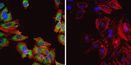
![ICC/IF analysis of HeLa cells using GTX25478 HSP60 antibody [4B9/89]. Cells were probed without (right) or with(left) an antibody. Green : Primary antibody Blue : Nuclei Red : Actin Fixation : formaldehyde Dilution : 1:100 overnight at 4 oC](https://www.genetex.com/upload/website/prouct_img/normal/GTX25478/GTX25478_665_ICC-IF_w_23060722_963.webp "ICC/IF analysis of HeLa cells using GTX25478 HSP60 antibody [4B9/89]. Cells were probed without (right) or with(left) an antibody. Green : Primary antibody Blue : Nuclei Red : Actin Fixation : formaldehyde Dilution : 1:100 overnight at 4 oC")
![FACS analysis of permeabilized, fixed HeLa cells using GTX25478 HSP60 antibody [4B9/89]. Orange histogram : Primary antibody Black histogram : Isotype control antibody Dilution : 10 μg/ml incubated for 1 hour on ice](https://www.genetex.com/upload/website/prouct_img/normal/GTX25478/GTX25478_176_FACS_w_23060722_874.webp "FACS analysis of permeabilized, fixed HeLa cells using GTX25478 HSP60 antibody [4B9/89]. Orange histogram : Primary antibody Black histogram : Isotype control antibody Dilution : 10 μg/ml incubated for 1 hour on ice")
![ICC/IF analysis of human endothelial cells using GTX25478 HSP60 antibody [4B9/89].](https://www.genetex.com/upload/website/prouct_img/normal/GTX25478/GTX25478_662_ICC-IF_w_23060722_232.webp "ICC/IF analysis of human endothelial cells using GTX25478 HSP60 antibody [4B9/89].")
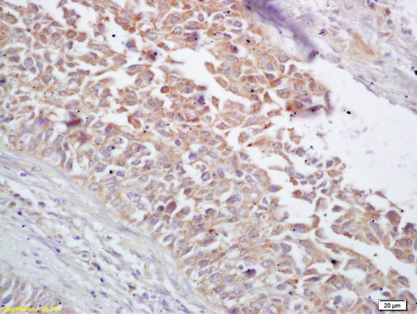
![IHC-P analysis of human breast carcinoma tissue using GTX25478 HSP60 antibody [4B9/89]. Left : Primary antibody Right : Negative control without primary antibody Antigen retrieval : heat induced antigen retrieval was performed using 10mM sodium citrate (pH6.0) buffer, microwaved for 8-15 minutes Dilution : 1:50](https://www.genetex.com/upload/website/prouct_img/normal/GTX25478/GTX25478_1292_IHC-P_w_23060722_302.webp "IHC-P analysis of human breast carcinoma tissue using GTX25478 HSP60 antibody [4B9/89]. Left : Primary antibody Right : Negative control without primary antibody Antigen retrieval : heat induced antigen retrieval was performed using 10mM sodium citrate (pH6.0) buffer, microwaved for 8-15 minutes Dilution : 1:50")
![IHC-P analysis of human kidney tissue using GTX25478 HSP60 antibody [4B9/89]. Left : Primary antibody Right : Negative control without primary antibody Antigen retrieval : heat induced antigen retrieval was performed using 10mM sodium citrate (pH6.0) buffer, microwaved for 8-15 minutes Dilution : 1:100](https://www.genetex.com/upload/website/prouct_img/normal/GTX25478/GTX25478_1293_IHC-P_w_23060722_862.webp "IHC-P analysis of human kidney tissue using GTX25478 HSP60 antibody [4B9/89]. Left : Primary antibody Right : Negative control without primary antibody Antigen retrieval : heat induced antigen retrieval was performed using 10mM sodium citrate (pH6.0) buffer, microwaved for 8-15 minutes Dilution : 1:100")
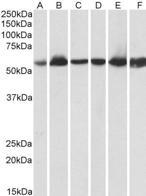
![WB analysis of 50μg of the indicated whole cell lysates using GTX25478 HSP60 antibody [4B9/89]. Dilution : 1:1000](https://www.genetex.com/upload/website/prouct_img/normal/GTX25478/GTX25478_1782_WB_w_23060722_184.webp "WB analysis of 50μg of the indicated whole cell lysates using GTX25478 HSP60 antibody [4B9/89]. Dilution : 1:1000")
![ICC/IF analysis of ATDC5 Cells using GTX25478 HSP60 antibody [4B9/89]. Cells were probed without (right) or with(left) an antibody. Green : Primary antibody Blue : Nuclei Red : Actin Fixation : formaldehyde Dilution : 1:100 overnight at 4 oC](https://www.genetex.com/upload/website/prouct_img/normal/GTX25478/GTX25478_664_ICC-IF_w_23060722_476.webp "ICC/IF analysis of ATDC5 Cells using GTX25478 HSP60 antibody [4B9/89]. Cells were probed without (right) or with(left) an antibody. Green : Primary antibody Blue : Nuclei Red : Actin Fixation : formaldehyde Dilution : 1:100 overnight at 4 oC")
![ICC/IF analysis of A2058 Cells using GTX25478 HSP60 antibody [4B9/89]. Cells were probed without (right) or with(left) an antibody. Green : Primary antibody Blue : Nuclei Red : Actin Fixation : formaldehyde Dilution : 1:200 overnight at 4oC](https://www.genetex.com/upload/website/prouct_img/normal/GTX25478/GTX25478_663_ICC-IF_w_23060722_781.webp "ICC/IF analysis of A2058 Cells using GTX25478 HSP60 antibody [4B9/89]. Cells were probed without (right) or with(left) an antibody. Green : Primary antibody Blue : Nuclei Red : Actin Fixation : formaldehyde Dilution : 1:200 overnight at 4oC")
ICC/IF analysis of HeLa cells using GTX25478 HSP60 antibody [4B9/89]. Cells were probed without (right) or with(left) an antibody. Green : Primary antibody Blue : Nuclei Red : Actin Fixation : Formalin Permeabilization : 0.1% Triton X-100 in TBS for 10 minutes Dilution : 1:50 for at least 1 hour at room temperature
HSP60 antibody [4B9/89]
GTX25478
ApplicationsFlow Cytometry, ImmunoFluorescence, ImmunoPrecipitation, Western Blot, ELISA, ImmunoCytoChemistry, ImmunoHistoChemistry, ImmunoHistoChemistry Paraffin, Neutralisation/Blocking
Product group Antibodies
ReactivityBacteria, Human, Mouse, Primate, Rat
TargetHSPD1
Overview
- SupplierGeneTex
- Product NameHSP60 antibody [4B9/89]
- Delivery Days Customer9
- Application Supplier NoteWB: 1:100 - 1:1000. ICC/IF: 10 microg/ml. IHC-P: 1:200. FCM: 1-20 microg/ml. IP: 2microg. *Optimal dilutions/concentrations should be determined by the researcher.Not tested in other applications.
- ApplicationsFlow Cytometry, ImmunoFluorescence, ImmunoPrecipitation, Western Blot, ELISA, ImmunoCytoChemistry, ImmunoHistoChemistry, ImmunoHistoChemistry Paraffin, Neutralisation/Blocking
- CertificationResearch Use Only
- ClonalityMonoclonal
- Clone ID4B9/89
- ConjugateUnconjugated
- Gene ID3329
- Target nameHSPD1
- Target descriptionheat shock protein family D (Hsp60) member 1
- Target synonymsCPN60, GROEL, HLD4, HSP-60, HSP60, HSP65, HuCHA60, SPG13, 60 kDa heat shock protein, mitochondrial, 60 kDa chaperonin, P60 lymphocyte protein, chaperonin 60, epididymis secretory sperm binding protein, heat shock 60kDa protein 1 (chaperonin), heat shock protein 65, heat shock protein family D member 1, mitochondrial matrix protein P1, short heat shock protein 60 Hsp60s1
- HostMouse
- IsotypeIgG2a
- Protein IDP10809
- Protein Name60 kDa heat shock protein, mitochondrial
- Scientific DescriptionThis gene encodes a member of the chaperonin family. The encoded mitochondrial protein may function as a signaling molecule in the innate immune system. This protein is essential for the folding and assembly of newly imported proteins in the mitochondria. This gene is adjacent to a related family member and the region between the 2 genes functions as a bidirectional promoter. Several pseudogenes have been associated with this gene. Two transcript variants encoding the same protein have been identified for this gene. Mutations associated with this gene cause autosomal recessive spastic paraplegia 13. [provided by RefSeq, Jun 2010]
- ReactivityBacteria, Human, Mouse, Primate, Rat
- Storage Instruction-20°C or -80°C,2°C to 8°C
- UNSPSC41116161
References
- Overexpression of the mitochondrial pyruvate carrier reduces lactate production and increases recombinant protein productivity in CHO cells. Bulte DB et al., 2020 Sep, Biotechnol BioengRead this paper
Datasheet
Related products
Product group Antibodies
Anti-Hsp60 AntibodyA83554
ApplicationsImmunoFluorescence, Western Blot, ELISA, ImmunoHistoChemistry
ReactivityHuman, Porcine, Rat
- SizePrice
Product group Antibodies
Anti-HSP60 Antibody130-10057
ApplicationsELISA
ReactivityHuman
TargetHSPD1
- SizePrice
Product group Antibodies
References
HSP60 Polyclonal AntibodyBS-0191R
ApplicationsImmunoFluorescence, Western Blot, ELISA, ImmunoCytoChemistry, ImmunoHistoChemistry, ImmunoHistoChemistry Frozen, ImmunoHistoChemistry Paraffin
ReactivityBovine, Canine, Equine, Human, Mouse, Rabbit, Rat
TargetHSPD1
- SizePrice
Product group Antibodies
HSPD1 AntibodyCSB-PA002990
ApplicationsImmunoFluorescence, Western Blot, ELISA, ImmunoHistoChemistry
ReactivityHuman, Mouse, Rat
TargetHSPD1
- SizePrice
Product group Antibodies

ApplicationsImmunoFluorescence, Western Blot, ELISA, ImmunoHistoChemistry
ReactivityBovine, Canine, Human, Porcine, Rat
TargetHSPD1
- SizePrice
Product group Antibodies
ApplicationsFlow Cytometry
TargetHSPD1
- SizePrice
![ICC/IF analysis of A431 cells using GTX25479 HSP60 antibody [2E1/53]. Cells were probed without (right) or with(left) an antibody. Green : Primary antibody Blue : Nuclei Red : Actin Fixation : Formalin Permeabilization : 0.1% Triton X-100 in TBS for 5-10 minute Dilution : 1:100 incubated overnight in a humidified chamber](https://www.genetex.com/upload/website/prouct_img/normal/GTX25479/GTX25479_668_ICC-IF_w_23060722_504.webp)
Product group Antibodies
HSP60 antibody [2E1/53]GTX25479
ApplicationsImmunoFluorescence, ImmunoPrecipitation, Western Blot, ELISA, ImmunoCytoChemistry, ImmunoHistoChemistry, ImmunoHistoChemistry Paraffin
ReactivityHuman, Mouse, Primate, Rat
TargetHSPD1
- SizePrice
Product group Antibodies

ApplicationsELISA
ReactivityHuman
TargetHSPD1
- SizePrice
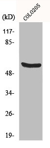
![WB analysis of HeLa whole cell lysate using GTX02649 HSP60 antibody [HSPD1/2206R].](https://www.genetex.com/upload/website/prouct_img/normal/GTX02649/GTX02649_20210319_WB_w_23053122_319.webp)
Product group Antibodies
HSP60 antibody [HSPD1/2206R]GTX02649
ApplicationsWestern Blot, ImmunoHistoChemistry, ImmunoHistoChemistry Paraffin
ReactivityBovine, Canine, Chicken, Hamster, Human, Monkey, Mouse, Porcine, Rabbit, Rat, Sheep
TargetHSPD1
- SizePrice

Product group Antibodies
Anti-HSPD1 AntibodyHPA001523
ApplicationsWestern Blot, ImmunoCytoChemistry, ImmunoHistoChemistry
ReactivityHuman, Mouse, Rat
TargetHSPD1
- SizePrice