

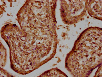
IHC image of CSB-PA835695LA01HU diluted at 1:500 and staining in paraffin-embedded human kidney tissue performed on a Leica BondTM system. After dewaxing and hydration, antigen retrieval was mediated by high pressure in a citrate buffer (pH 6.0). Section was blocked with 10% normal goat serum 30min at RT. Then primary antibody (1% BSA) was incubated at 4°C overnight. The primary is detected by a biotinylated secondary antibody and visualized using an HRP conjugated SP system.
. Section was blocked with 10% normal goat serum 30min at RT. Then primary antibody (1% BSA) was incubated at 4°C overnight. The primary is detected by a biotinylated secondary antibody and visualized using an HRP conjugated SP system.")
.")
IHC image of CSB-PA835695LA01HU diluted at 1:500 and staining in paraffin-embedded human kidney tissue performed on a Leica BondTM system. After dewaxing and hydration, antigen retrieval was mediated by high pressure in a citrate buffer (pH 6.0). Section was blocked with 10% normal goat serum 30min at RT. Then primary antibody (1% BSA) was incubated at 4°C overnight. The primary is detected by a biotinylated secondary antibody and visualized using an HRP conjugated SP system.
HTRA1 Antibody
CSB-PA835695LA01HU
ApplicationsImmunoFluorescence, ELISA, ImmunoHistoChemistry
Product group Antibodies
ReactivityHuman
TargetHTRA1
Overview
- SupplierCusabio
- Product NameHTRA1 Antibody
- Delivery Days Customer20
- ApplicationsImmunoFluorescence, ELISA, ImmunoHistoChemistry
- CertificationResearch Use Only
- ClonalityPolyclonal
- ConjugateUnconjugated
- Gene ID5654
- Target nameHTRA1
- Target descriptionHtrA serine peptidase 1
- Target synonymsARMD7, CADASIL2, CARASIL, CARASIL2, HtrA, L56, ORF480, PRSS11, serine protease HTRA1, IGFBP5-protease, high-temperature requirement A serine peptidase 1, protease, serine, 11 (IGF binding)
- HostRabbit
- IsotypeIgG
- Protein IDQ92743
- Protein NameSerine protease HTRA1
- Scientific DescriptionSerine protease with a variety of targets, including extracellular matrix proteins such as fibronectin. HTRA1-generated fibronectin fragments further induce synovial cells to up-regulate MMP1 and MMP3 production. May also degrade proteoglycans, such as aggrecan, decorin and fibromodulin. Through cleavage of proteoglycans, may release soluble FGF-glycosaminoglycan complexes that promote the range and intensity of FGF signals in the extracellular space. Regulates the availability of insulin-like growth factors (IGFs) by cleaving IGF-binding proteins. Inhibits signaling mediated by TGF-beta family members. This activity requires the integrity of the catalytic site, although it is unclear whether TGF-beta proteins are themselves degraded. By acting on TGF-beta signaling, may regulate many physiological processes, including retinal angiogenesis and neuronal survival and maturation during development. Intracellularly, degrades TSC2, leading to the activation of TSC2 downstream targets.
- ReactivityHuman
- Storage Instruction-20°C or -80°C
- UNSPSC41116161
Related products
Product group Antibodies

HTRA1 AntibodyLS-C832624
ApplicationsWestern Blot, ELISA, ImmunoHistoChemistry
ReactivityHuman, Mouse, Rat
TargetHTRA1
- SizePrice

Product group Antibodies
Anti-HTRA1 Antibody Picoband(r)A01801-1-CARRIER-FREE
ApplicationsWestern Blot, ImmunoHistoChemistry
ReactivityHuman, Mouse, Rat
TargetHTRA1
- SizePrice
Product group Antibodies
HTRA1 Recombinant AntibodyBSM-62786R
ApplicationsImmunoFluorescence, Western Blot, ImmunoCytoChemistry
ReactivityHuman
TargetHTRA1
- SizePrice
Product group Antibodies
ApplicationsImmunoPrecipitation, Western Blot, ImmunoCytoChemistry, ImmunoHistoChemistry
ReactivityPorcine
TargetHTRA1
- SizePrice
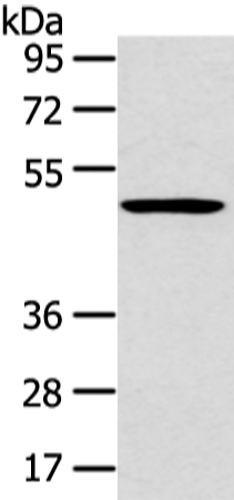
![Various whole cell extracts (30 μg) were separated by 10% SDS-PAGE, and the membrane was blotted with HtrA1 antibody [HL3709] (GTX641881) diluted at 1:1000. The HRP-conjugated anti-rabbit IgG antibody (GTX213110-01) was used to detect the primary antibody, and the signal was developed with Trident ECL plus-Enhanced. Corresponding RNA expression data are based on Human Protein Atlas program.](https://www.genetex.com/upload/website/prouct_img/normal/GTX641881/GTX641881_45726_20250328_WB_TPM_watermark_25040100_413.webp)
Product group Antibodies
HtrA1 antibody [HL3709]GTX641881
ApplicationsWestern Blot
ReactivityHuman
TargetHTRA1
- SizePrice

Product group Antibodies
Anti-HTRA1 AntibodyHPA036655
ApplicationsImmunoCytoChemistry
ReactivityHuman
TargetHTRA1
- SizePrice