

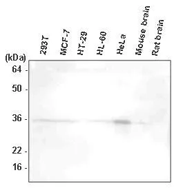
WB analysis of 293T, MCF7, HT-29, HL-60, HeLa, mouse brain, and rat brain lysates using HtrA2/Omi antibody at a dilution of 1:1,000.
WB analysis of 293T, MCF7, HT-29, HL-60, HeLa, mouse brain, and rat brain lysates using HtrA2/Omi antibody at a dilution of 1:1,000.
HTRA2 antibody [1B3]
GTX53729
ApplicationsWestern Blot, ELISA
Product group Antibodies
ReactivityHuman, Mouse
TargetHTRA2
Overview
- SupplierGeneTex
- Product NameHTRA2 antibody [1B3]
- Delivery Days Customer9
- Application Supplier NoteThe antibody has been tested by ELISA and Western blot analysis to assure specificity and reactivity. Since application varies, however, each investigation should be titrated by the reagent to obtain optimal results. Recommended dilution range for Western blot analysis is 1:500 ~ 2,000. Recommended starting dilution is 1:1,000.
- ApplicationsWestern Blot, ELISA
- CertificationResearch Use Only
- ClonalityMonoclonal
- Clone ID1B3
- Concentration1 mg/ml
- ConjugateUnconjugated
- Gene ID27429
- Target nameHTRA2
- Target descriptionHtrA serine peptidase 2
- Target synonymsMGCA8, OMI, PARK13, PRSS25, serine protease HTRA2, mitochondrial, HtrA-like serine protease, Omi stress-regulated endoprotease, epididymis secretory sperm binding protein, high temperature requirement protein A2, protease, serine, 25, serine protease 25, serine proteinase OMI
- HostMouse
- IsotypeIgG1
- Protein IDO43464
- Protein NameSerine protease HTRA2, mitochondrial
- Scientific DescriptionThis gene encodes a serine protease. The protein has been localized in the endoplasmic reticulum and interacts with an alternatively spliced form of mitogen-activated protein kinase 14. The protein has also been localized to the mitochondria with release to the cytosol following apoptotic stimulus. The protein is thought to induce apoptosis by binding the apoptosis inhibitory protein baculoviral IAP repeat-containing 4. Nuclear localization of this protein has also been observed. Alternate splicing of this gene results in multiple transcript variants encoding different isoforms. [provided by RefSeq, Mar 2016]
- ReactivityHuman, Mouse
- Storage Instruction-20°C or -80°C,2°C to 8°C
- UNSPSC41116161
Datasheet
Related products
Product group Antibodies
Phospho-HTRA2 (S212) AntibodyCSB-PA008882
ApplicationsELISA, ImmunoHistoChemistry
ReactivityHuman, Mouse, Rat
TargetHTRA2
- SizePrice

Product group Antibodies
Anti-HTRA2 Antibody Picoband(r)A01941-3-CARRIER-FREE
ApplicationsFlow Cytometry, ImmunoFluorescence, Western Blot, ELISA, ImmunoCytoChemistry, ImmunoHistoChemistry
ReactivityHuman
TargetHTRA2
- SizePrice
Product group Antibodies
Anti-HTRA2 AntibodyA31033
ApplicationsImmunoFluorescence, Western Blot, ImmunoHistoChemistry
ReactivityHuman, Mouse, Rat
- SizePrice
Product group Antibodies

HTRA2 / OMI Antibody (Preservative Free)LS-C149197
ApplicationsELISA
ReactivityHuman
TargetHTRA2
- SizePrice

Product group Antibodies
Anti-HTRA2 AntibodyHPA006602
ApplicationsImmunoCytoChemistry
ReactivityHuman
TargetHTRA2
- SizePrice
Product group Antibodies
HTRA2 Polyclonal AntibodyCAC14730
ApplicationsImmunoPrecipitation, Western Blot, ELISA, ImmunoHistoChemistry
TargetHTRA2
- SizePrice
Product group Antibodies
HTRA2 antibodyGTX110703
ApplicationsWestern Blot, ImmunoHistoChemistry, ImmunoHistoChemistry Paraffin
ReactivityHuman
TargetHTRA2
- SizePrice
Product group Antibodies
HTRA2 antibodyGTX132456
ApplicationsWestern Blot
ReactivityHuman
TargetHTRA2
- SizePrice