

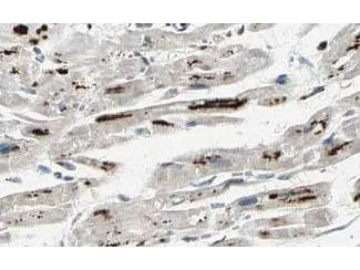
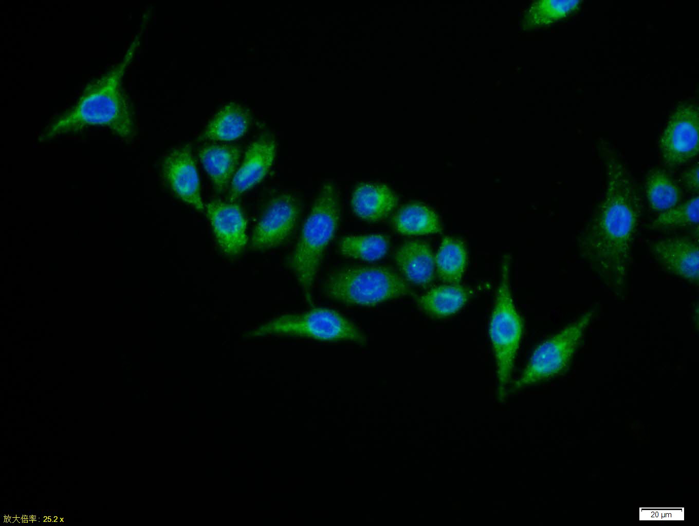
GeneTex's Affinity Purified anti-iASPP antibody shows strong cytoplasmic and membranous staining of myocytes in human heart tissue. Tissue was formalin-fixed and paraffin embedded. Brown color indicates presence of protein, blue color shows cell nuclei.
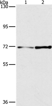
![Western blot using GeneTex's affinity purified anti-iASPP antibody shows detection of a band at ~100 kDa (arrowhead) corresponding to isoform 1 of iASPP in MCF7 whole cell lysates. Preincubation with immunizing peptide blocks specific band staining (data not shown). Approximately 35 μg of lysate was separated by 4-20% Tris Glycine SDS-PAGE. After blocking, the membrane was probed with the primary antibody diluted to 1:1,500 in 5% BLOTTO/PBS overnight at 4oC. The membrane was washed and reacted with a 1:10,000 dilution of IRDye800 conjugated goat anti-Rabbit IgG [H&L] for 45 min at room temperature (800 nm channel, green). Molecular weight estimation was made by comparison to prestained MW markers. IRDye800 fluorescence image was captured using the OdysseyR Infrared Imaging System developed by LI-COR. IRDye is a trademark of LI-COR, Inc. Other detection systems will yield similar results.](https://www.genetex.com/upload/website/prouct_img/normal/GTX48696/GTX48696_20160330_WB_w_23060823_143.webp "Western blot using GeneTex's affinity purified anti-iASPP antibody shows detection of a band at ~100 kDa (arrowhead) corresponding to isoform 1 of iASPP in MCF7 whole cell lysates. Preincubation with immunizing peptide blocks specific band staining (data not shown). Approximately 35 μg of lysate was separated by 4-20% Tris Glycine SDS-PAGE. After blocking, the membrane was probed with the primary antibody diluted to 1:1,500 in 5% BLOTTO/PBS overnight at 4oC. The membrane was washed and reacted with a 1:10,000 dilution of IRDye800 conjugated goat anti-Rabbit IgG [H&L] for 45 min at room temperature (800 nm channel, green). Molecular weight estimation was made by comparison to prestained MW markers. IRDye800 fluorescence image was captured using the OdysseyR Infrared Imaging System developed by LI-COR. IRDye is a trademark of LI-COR, Inc. Other detection systems will yield similar results.")

GeneTex's Affinity Purified anti-iASPP antibody shows strong cytoplasmic and membranous staining of myocytes in human heart tissue. Tissue was formalin-fixed and paraffin embedded. Brown color indicates presence of protein, blue color shows cell nuclei.
Iaspp antibody
GTX48696
ApplicationsWestern Blot, ELISA, ImmunoHistoChemistry, ImmunoHistoChemistry Paraffin
Product group Antibodies
ReactivityHuman
TargetPPP1R13L
Overview
- SupplierGeneTex
- Product NameIaspp antibody
- Delivery Days Customer9
- Application Supplier NoteWB: 1:1000-1:5000. IHC-P: 1:1000-1:5000. ELISA: 1:40000-1:160000. *Optimal dilutions/concentrations should be determined by the researcher.Not tested in other applications.
- ApplicationsWestern Blot, ELISA, ImmunoHistoChemistry, ImmunoHistoChemistry Paraffin
- CertificationResearch Use Only
- ClonalityPolyclonal
- Concentration1.1 mg/ml
- ConjugateUnconjugated
- Gene ID10848
- Target namePPP1R13L
- Target descriptionprotein phosphatase 1 regulatory subunit 13 like
- Target synonymsARCME, CMAEA, IASPP, NKIP1, RAI, RAI4, relA-associated inhibitor, NFkB interacting protein 1, PPP1R13B-like protein, inhibitor of ASPP protein, inhibitor of apoptosis stimulating protein of p53, protein iASPP, protein phosphatase 1, regulatory (inhibitor) subunit 13 like, retinoic acid induced 4
- HostRabbit
- IsotypeIgG
- Protein IDQ8WUF5
- Protein NameRelA-associated inhibitor
- Scientific DescriptionASPP proteins (ASPP1, ASPP2 and iASPP) represent a new family of p53 binding proteins. ASPP1 and ASPP2 bind and enhance p53-mediated apoptosis. In contrast, the third member, iASPP, functionally inactivates p53. iASPP (also called protein phosphatase 1 regulatory (inhibitor) subunit 13 like protein, Inhibitor of ASPP protein, Protein iASPP, PPP1R13B-like protein and NFkB-interacting protein 1) plays a central role in regulation of apoptosis and transcription via its interaction with NF-kappa-B and p53/TP53 proteins. iASPP blocks transcription of HIV-1 virus by inhibiting the action of both NF-kappa-B and SP1. This protein
- ReactivityHuman
- Storage Instruction-20°C or -80°C,2°C to 8°C
- UNSPSC41116161
Datasheet
Related products
Product group Antibodies
PPP1R13L AntibodyCSB-PA290837
ApplicationsWestern Blot, ELISA, ImmunoHistoChemistry
ReactivityHuman, Mouse
TargetPPP1R13L
- SizePrice
Product group Antibodies
Anti-PPP1R13L AntibodyA36717
ApplicationsWestern Blot, ImmunoHistoChemistry
ReactivityHuman, Mouse
- SizePrice

Product group Antibodies
Anti-PPP1R13L AntibodyHPA041231
ApplicationsWestern Blot, ImmunoCytoChemistry, ImmunoHistoChemistry
ReactivityHuman
TargetPPP1R13L
- SizePrice
Product group Antibodies

PPP1R13L / iASPP AntibodyLS-C405660
ApplicationsWestern Blot, ELISA, ImmunoHistoChemistry
ReactivityHuman, Mouse
TargetPPP1R13L
- SizePrice
Product group Antibodies
IASPP antibodyGTX37659
ApplicationsWestern Blot, ImmunoHistoChemistry, ImmunoHistoChemistry Paraffin
ReactivityHuman, Mouse
TargetPPP1R13L
- SizePrice
Product group Antibodies
References
IASPP Polyclonal AntibodyBS-0284R
ApplicationsFlow Cytometry, ImmunoFluorescence, Western Blot, ELISA, ImmunoCytoChemistry, ImmunoHistoChemistry, ImmunoHistoChemistry Frozen, ImmunoHistoChemistry Paraffin
ReactivityBovine, Canine, Guinea Pig, Human, Mouse, Rat, Sheep
TargetPPP1R13L
- SizePrice
Product group Antibodies
Anti-PPP1R13L Antibody144-61893
ApplicationsWestern Blot
ReactivityHuman, Mouse
TargetPPP1R13L
- SizePrice