

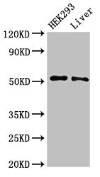
Western Blot Positive WB detected in: HEK293 whole cell lysate, Rat liver tissue All lanes: IFIT2 antibody at 3microg/ml Secondary Goat polyclonal to rabbit IgG at 1/50000 dilution Predicted band size: 55 kDa Observed band size: 55 kDa
. Section was blocked with 10% normal goat serum 30min at RT. Then primary antibody (1% BSA) was incubated at 4°C overnight. The primary is detected by a biotinylated secondary antibody and visualized using an HRP conjugated SP system.")
. Section was blocked with 10% normal goat serum 30min at RT. Then primary antibody (1% BSA) was incubated at 4°C overnight. The primary is detected by a biotinylated secondary antibody and visualized using an HRP conjugated SP system.")
")
Western Blot Positive WB detected in: HEK293 whole cell lysate, Rat liver tissue All lanes: IFIT2 antibody at 3microg/ml Secondary Goat polyclonal to rabbit IgG at 1/50000 dilution Predicted band size: 55 kDa Observed band size: 55 kDa
IFIT2 Antibody
CSB-PA011021LA01HU
ApplicationsImmunoFluorescence, Western Blot, ELISA, ImmunoHistoChemistry
Product group Antibodies
ReactivityHuman, Rat
TargetIFIT2
Overview
- SupplierCusabio
- Product NameIFIT2 Antibody
- Delivery Days Customer20
- ApplicationsImmunoFluorescence, Western Blot, ELISA, ImmunoHistoChemistry
- CertificationResearch Use Only
- ClonalityPolyclonal
- ConjugateUnconjugated
- Gene ID3433
- Target nameIFIT2
- Target descriptioninterferon induced protein with tetratricopeptide repeats 2
- Target synonymsG10P2, GARG-39, IFI-54, IFI-54K, IFI54, IFIT-2, ISG-54, ISG-54 K, ISG-54K, ISG54, P54, cig42, interferon-induced protein with tetratricopeptide repeats 2, Interferon, alpha-inducible protein (MW 54kD), interferon-induced 54 kDa protein, interferon-induced protein 54
- HostRabbit
- IsotypeIgG
- Protein IDP09913
- Protein NameInterferon-induced protein with tetratricopeptide repeats 2
- Scientific DescriptionIFN-induced antiviral protein which inhibits expression of viral messenger RNAs lacking 2-O-methylation of the 5 cap. The ribose 2-O-methylation would provide a molecular signature to distinguish between self and non-self mRNAs by the host during viral infection. Viruses evolved several ways to evade this restriction system such as encoding their own 2-O-methylase for their mRNAs or by stealing host cap containing the 2-O-methylation (cap snatching mechanism). Binds AU-rich viral RNAs, with or without 5 triphosphorylation, RNA-binding is required for antiviral activity. Can promote apoptosis.
- ReactivityHuman, Rat
- Storage Instruction-20°C or -80°C
- UNSPSC41116161
Related products
Product group Antibodies

ISG54 / IFIT2 AntibodyLS-C748789
ApplicationsWestern Blot
ReactivityHuman
TargetIFIT2
- SizePrice

Product group Antibodies
Anti-IFIT2 AntibodyHPA003408
ApplicationsImmunoHistoChemistry
ReactivityHuman
TargetIFIT2
- SizePrice
Product group Antibodies
IFIT2 Polyclonal AntibodyCAC14848
ApplicationsImmunoFluorescence, Western Blot, ELISA, ImmunoHistoChemistry
ReactivityRat
TargetIFIT2
- SizePrice
![IFIT2 antibody [N1], N-term detects IFIT2 protein at endoplasmic reticulum by immunofluorescent analysis. Sample: HeLa cells were fixed in 4% paraformaldehyde at RT for 15 min. Green: IFIT2 protein stained by IFIT2 antibody [N1], N-term (GTX108346) diluted at 1:200. Red: alpha Tubulin, a cytoskeleton marker, stained by alpha Tubulin antibody [B-5-1-2] (GTX11304) diluted at 1:10000. Blue: Hoechst 33342 staining.](https://www.genetex.com/upload/website/prouct_img/normal/GTX108346/GTX108346_39904_20150410_IFA_w_23060120_406.webp)
Product group Antibodies
IFIT2 antibody [N1], N-termGTX108346
ApplicationsImmunoFluorescence, Western Blot, ImmunoCytoChemistry
ReactivityHuman
TargetIFIT2
- SizePrice
Product group Antibodies
Anti-IFIT2Y058825
ApplicationsWestern Blot, ImmunoHistoChemistry
ReactivityHuman
- SizePrice
Product group Antibodies
Anti-IFIT2 Antibody144-62897
ApplicationsWestern Blot
ReactivityHuman
TargetIFIT2
- SizePrice
Product group Antibodies
IFIT2 Polyclonal AntibodyBS-15528R
ApplicationsImmunoFluorescence, ELISA, ImmunoCytoChemistry, ImmunoHistoChemistry, ImmunoHistoChemistry Frozen, ImmunoHistoChemistry Paraffin
ReactivityBovine, Human, Mouse, Porcine, Rat, Sheep
- SizePrice