

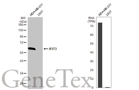
Various whole cell extracts (30 μg) were separated by 10% SDS-PAGE, and the membrane was blotted with IFIT3 antibody [HL2468] (GTX638817) diluted at 1:1000. The HRP-conjugated anti-rabbit IgG antibody (GTX213110-01) was used to detect the primary antibody. Corresponding RNA expression data for the same cell lines are based on Human Protein Atlas program.
![IFIT3 antibody [HL2468] detects IFIT3 protein at mitochondria by immunohistochemical analysis. Sample: Paraffin-embedded human glioblastoma. IFIT3 stained by IFIT3 antibody [HL2468] (GTX638817) diluted at 1:100. Antigen Retrieval: Citrate buffer, pH 6.0, 15 min](https://www.genetex.com/upload/website/prouct_img/normal/GTX638817/GTX638817_T-45096_20230721_IHC-P_23073119_274.webp "IFIT3 antibody [HL2468] detects IFIT3 protein at mitochondria by immunohistochemical analysis. Sample: Paraffin-embedded human glioblastoma. IFIT3 stained by IFIT3 antibody [HL2468] (GTX638817) diluted at 1:100. Antigen Retrieval: Citrate buffer, pH 6.0, 15 min")
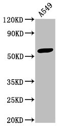
![Untreated (–) and treated (+) A549 whole cell extracts (50 μg) were separated by 10% SDS-PAGE, and the membrane was blotted with IFIT3 antibody [HL2468] (GTX638817) diluted at 1:1000. The HRP-conjugated anti-rabbit IgG antibody (GTX213110-01) was used to detect the primary antibody.](https://www.genetex.com/upload/website/prouct_img/normal/GTX638817/GTX638817_45159_20230908_WB_treatment_IFN-gamma_23091319_604.webp "Untreated (–) and treated (+) A549 whole cell extracts (50 μg) were separated by 10% SDS-PAGE, and the membrane was blotted with IFIT3 antibody [HL2468] (GTX638817) diluted at 1:1000. The HRP-conjugated anti-rabbit IgG antibody (GTX213110-01) was used to detect the primary antibody.")
![IFIT3 antibody [HL2468] detects IFIT3 protein at cytoplasm by immunofluorescent analysis. Sample: MDA-MB-231 cells were fixed in ice-cold MeOH for 5 min. Green: IFIT3 stained by IFIT3 antibody [HL2468] (GTX638817) diluted at 1:500. Blue: Fluoroshield with DAPI (GTX30920).](https://www.genetex.com/upload/website/prouct_img/normal/GTX638817/GTX638817_T-45096_20230908_ICC_IF_23091901_989.webp "IFIT3 antibody [HL2468] detects IFIT3 protein at cytoplasm by immunofluorescent analysis. Sample: MDA-MB-231 cells were fixed in ice-cold MeOH for 5 min. Green: IFIT3 stained by IFIT3 antibody [HL2468] (GTX638817) diluted at 1:500. Blue: Fluoroshield with DAPI (GTX30920).")
Various whole cell extracts (30 μg) were separated by 10% SDS-PAGE, and the membrane was blotted with IFIT3 antibody [HL2468] (GTX638817) diluted at 1:1000. The HRP-conjugated anti-rabbit IgG antibody (GTX213110-01) was used to detect the primary antibody. Corresponding RNA expression data for the same cell lines are based on Human Protein Atlas program.
IFIT3 antibody [HL2468]
GTX638817
ApplicationsImmunoFluorescence, Western Blot, ImmunoCytoChemistry, ImmunoHistoChemistry, ImmunoHistoChemistry Paraffin
Product group Antibodies
ReactivityHuman
TargetIFIT3
Overview
- SupplierGeneTex
- Product NameIFIT3 antibody [HL2468]
- Delivery Days Customer9
- Application Supplier NoteWB: 1:500-1:3000. *Optimal dilutions/concentrations should be determined by the researcher.Not tested in other applications.
- ApplicationsImmunoFluorescence, Western Blot, ImmunoCytoChemistry, ImmunoHistoChemistry, ImmunoHistoChemistry Paraffin
- CertificationResearch Use Only
- ClonalityMonoclonal
- Clone IDHL246
- Concentration1 mg/ml
- ConjugateUnconjugated
- Gene ID3437
- Target nameIFIT3
- Target descriptioninterferon induced protein with tetratricopeptide repeats 3
- Target synonymsCIG-49, GARG-49, IFI60, IFIT4, IRG2, ISG60, P60, RIG-G, cig41, interferon-induced protein with tetratricopeptide repeats 3, CIG49, IFI-60K, IFIT-3, IFIT-4, ISG-60, interferon-induced 60 kDa protein, interferon-induced protein with tetratricopeptide repeats 4, retinoic acid-induced gene G protein
- HostRabbit
- IsotypeIgG
- Protein IDO14879
- Protein NameInterferon-induced protein with tetratricopeptide repeats 3
- ReactivityHuman
- Storage Instruction-20°C or -80°C,2°C to 8°C
- UNSPSC41116161
Datasheet
Related products
Product group Antibodies
IFIT3 AntibodyCSB-PA011022HA01HU
ApplicationsImmunoFluorescence, ImmunoPrecipitation, Western Blot, ELISA, ImmunoHistoChemistry
ReactivityHuman
TargetIFIT3
- SizePrice
Product group Antibodies
Ifit3 Polyclonal AntibodyCAC07359
ApplicationsImmunoFluorescence, ImmunoPrecipitation, Western Blot, ELISA, ImmunoHistoChemistry
TargetIFIT3
- SizePrice
Product group Antibodies

IFIT3 AntibodyLS-C748372
ApplicationsWestern Blot
ReactivityHuman, Mouse
TargetIFIT3
- SizePrice

Product group Antibodies
Anti-IFIT3 AntibodyHPA059914
ApplicationsWestern Blot, ImmunoCytoChemistry, ImmunoHistoChemistry
ReactivityHuman
TargetIFIT3
- SizePrice
Product group Antibodies
Anti-IFIT3 AntibodyA03920-1
ApplicationsImmunoFluorescence, Western Blot, ImmunoHistoChemistry
ReactivityHuman, Mouse, Rat
TargetIFIT3
- SizePrice
Product group Antibodies
IFIT3 Polyclonal AntibodyBS-15515R
ApplicationsImmunoFluorescence, Western Blot, ELISA, ImmunoCytoChemistry, ImmunoHistoChemistry, ImmunoHistoChemistry Frozen, ImmunoHistoChemistry Paraffin
ReactivityHuman, Mouse, Rat
- SizePrice
Product group Antibodies
Anti-IFIT3 Antibody107-11025
ApplicationsImmunoFluorescence, Western Blot, ImmunoCytoChemistry, ImmunoHistoChemistry, ImmunoHistoChemistry Paraffin
ReactivityHuman
TargetIFIT3
- SizePrice
![IFIT3 antibody [HL2469] detects IFIT3 protein at mitochondria by immunohistochemical analysis. Sample: Paraffin-embedded human glioblastoma. IFIT3 stained by IFIT3 antibody [HL2469] (GTX638818) diluted at 1:100. Antigen Retrieval: Citrate buffer, pH 6.0, 15 min](https://www.genetex.com/upload/website/prouct_img/normal/GTX638818/GTX638818_T-45096_20230721_IHC-P_23073119_279.webp)
Product group Antibodies
IFIT3 antibody [HL2469]GTX638818
ApplicationsWestern Blot, ImmunoHistoChemistry, ImmunoHistoChemistry Paraffin
ReactivityHuman
TargetIFIT3
- SizePrice
![ICC/IF analysis of COS7 cells transiently transfected with IFIT3 plasmid using GTX84314 IFIT3 antibody [1G1].](https://www.genetex.com/upload/website/prouct_img/normal/GTX84314/GTX84314_1022_ICCIF_w_23061420_924.webp)
Product group Antibodies
References
IFIT3 antibody [1G1]GTX84314
ApplicationsFlow Cytometry, ImmunoFluorescence, Western Blot, ImmunoCytoChemistry
ReactivityHuman, Monkey
TargetIFIT3
- SizePrice