

IHC-plus(tm) DAPK3 / ZIP Kinase Antibody
LS-B557
ApplicationsImmunoFluorescence, Western Blot, ELISA, ImmunoCytoChemistry, ImmunoHistoChemistry, ImmunoHistoChemistry Paraffin
Product group Antibodies
ReactivityHuman, Mouse, Rat
TargetDAPK3
Overview
- SupplierLifeSpan BioSciences
- Product NameIHC-plus(tm) DAPK3 / ZIP Kinase Antibody
- Delivery Days Customer14
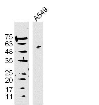
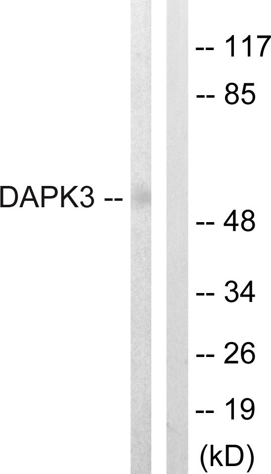
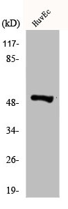
- Application Supplier NoteZIPK antibody can be used for detection of DNase II expression by Western blot at 1 ug/mL. An approximate 40 kDa band can be detected, which represents the pro-enzyme of DNase II. Antibody can also be used for immunocytochemistry starting at 5 ug/mL. For immunofluorescence start at 5 ug/mL. Antibody validated: Western Blot in human samples; Immunocytochemistry in human samples and Immunofluorescence in human samples. All other applications and species not yet tested. Western Blot: Predicted: 26 kDa Observed: 27 kDa. ELISA, ICC, IF, IHC, IHC-P (2.5 µg/ml), WB (1:500 - 1:1000) ZIPK antibody can be used for detection of DNase II expression by Western blot at 1 ug/mL. An approximate 40 kDa band can be detected, which represents the pro-enzyme of DNase II. Antibody can also be used for immunocytochemistry starting at 5 ug/mL. For immunofluorescence start at 5 ug/mL. Antibody validated: Western Blot in human samples; Immunocytochemistry in human samples and Immunofluorescence in human samples. All other applications and species not yet tested. Western Blot: Predicted: 26 kDa Observed: 27 kDa
- ApplicationsImmunoFluorescence, Western Blot, ELISA, ImmunoCytoChemistry, ImmunoHistoChemistry, ImmunoHistoChemistry Paraffin
- CertificationResearch Use Only
- ClonalityPolyclonal
- Concentration1 mg/ml
- ConjugateUnconjugated
- Gene ID1613
- Target nameDAPK3
- Target descriptiondeath associated protein kinase 3
- Target synonymsDLK, ZIP, ZIPK, death-associated protein kinase 3, DAP kinase 3, DAP-like kinase, MYPT1 kinase, ZIP-kinase, zipper-interacting protein kinase
- HostRabbit
- IsotypeIgG
- ReactivityHuman, Mouse, Rat
- Storage Instruction-20°C,2°C to 8°C
- UNSPSC41116161
Related products
Product group Antibodies
Anti-DAPK3 Antibody144-60895
ApplicationsWestern Blot
ReactivityHuman, Mouse, Rat
TargetDAPK3
- SizePrice

Product group Antibodies
References
ApplicationsImmunoFluorescence, Western Blot, ELISA, ImmunoHistoChemistry
ReactivityHuman, Mouse, Rat
TargetDAPK3
- SizePrice
![DAP Kinase 3 antibody detects DAP Kinase 3 protein at nucleus by immunofluorescent analysis. Sample: HeLa cells were fixed in 4% paraformaldehyde at RT for 15 min. Green: DAP Kinase 3 protein stained by DAP Kinase 3 antibody (GTX102404) diluted at 1:500. Red: alpha Tubulin, a cytoskeleton marker, stained by alpha Tubulin antibody [B-5-1-2] (GTX11304) diluted at 1:10000.](https://www.genetex.com/upload/website/prouct_img/normal/GTX102404/GTX102404_39820_20150410_IFA_w_23060100_953.webp)
Product group Antibodies
References
DAP Kinase 3 antibodyGTX102404
ApplicationsImmunoFluorescence, Western Blot, ImmunoCytoChemistry
ReactivityHuman, Mouse, Rat
TargetDAPK3
- SizePrice
Product group Antibodies
DAPK3 Polyclonal AntibodyCAC14638
ApplicationsImmunoFluorescence, Western Blot, ELISA
TargetDAPK3
- SizePrice
Product group Antibodies
ZIP Kinase Polyclonal Antibodybs-1692R
ApplicationsImmunoFluorescence, Western Blot, ELISA, ImmunoCytoChemistry, ImmunoHistoChemistry, ImmunoHistoChemistry Frozen, ImmunoHistoChemistry Paraffin
ReactivityBovine, Canine, Chicken, Equine, Human, Mouse, Rabbit, Rat
TargetDAPK3
- SizePrice
Product group Antibodies
Anti-DAPK3 AntibodyA95894
ApplicationsWestern Blot, ELISA, ImmunoHistoChemistry
ReactivityHuman, Mouse, Rat
- SizePrice
Product group Antibodies
DAPK3 AntibodyCSB-PA002072
ApplicationsWestern Blot, ELISA, ImmunoHistoChemistry
ReactivityHuman, Mouse, Rat
TargetDAPK3
- SizePrice
Product group Antibodies

DAPK3 / ZIP Kinase AntibodyLS-C750021
ApplicationsWestern Blot
ReactivityHuman, Mouse, Rat
TargetDAPK3
- SizePrice